Microwave-assisted preparation method of hydroxylapatite nanometer structure porous microspheres

A technology of hydroxyapatite and nanostructures, applied in chemical instruments and methods, phosphorus compounds, pharmaceutical formulations, etc., can solve the problems of increasing process complexity and production costs, and achieve good protein adsorption and drug loading and sustained release performance , simple process, environment-friendly appearance effect

Active Publication Date: 2013-05-22
SHANGHAI INST OF CERAMIC CHEM & TECH CHINESE ACAD OF SCI
View PDF8 Cites 27 Cited by
  • Summary
  • Abstract
  • Description
  • Claims
  • Application Information

AI Technical Summary

Problems solved by technology

The template added by this method needs to be cleaned in subsequent processing, thus increasing the complexity of the process and the production cost

Method used


Image

  • Microwave-assisted preparation method of hydroxylapatite nanometer structure porous microspheres
  • Microwave-assisted preparation method of hydroxylapatite nanometer structure porous microspheres
  • Microwave-assisted preparation method of hydroxylapatite nanometer structure porous microspheres

Examples


Embodiment 1

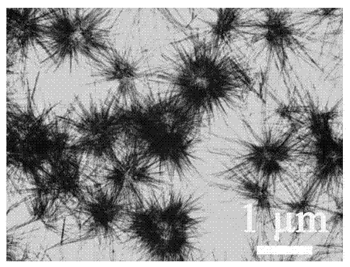
[0038] Mix 20mL of 4mg / L pyridoxal phosphate aqueous solution with 10mL of 0.05M calcium chloride aqueous solution, adjust the pH value of the mixed solution to 4.5 with ammonia water, place the mixed solution in a microwave reactor, and heat the microwave at a temperature of 180°C. After 5 minutes, the precipitate after the reaction was collected by centrifugation, washed with water, dried in air at 60° C., and then ground to obtain hollow microspheres assembled with hydroxyapatite nanorods.

[0039] The sample was characterized by TEM, such as figure 1 shown. It can be seen from the figure that the prepared sample structure is hollow microspheres assembled with nanorods, and the size of the microspheres is 0.5–2 μm.

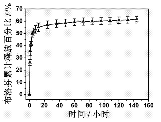
[0040] After the obtained sample was dried and ground, it was added to a solution of ibuprofen with a concentration of 40 mg / mL in n-hexane, and shaken for 24 hours at a rate of 160 rpm at 37°C. Then the sample was centrifuged, dried and ground at 60°C in air...

Embodiment 2

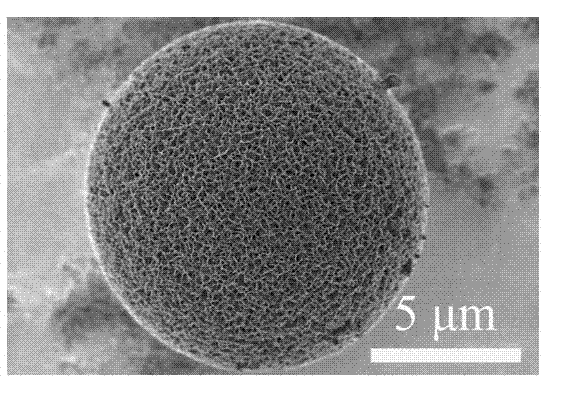
[0043] Mix 20mL of 4mg / L pyridoxal phosphate aqueous solution with 10mL of 0.05M calcium chloride aqueous solution, adjust the pH value of the mixed solution to 4.5 with ammonia water, place the mixed solution in a microwave reactor, and heat the microwave at a temperature of 140°C. After 5 minutes, the precipitate after the reaction was collected by centrifugation, washed with water, dried in air at 60° C., and then ground to obtain hydroxyapatite nanosheet-assembled microspheres.

[0044] The sample was characterized by SEM, such as image 3 shown. It can be seen from the figure that the prepared sample structure is nanosheet-assembled microspheres, and the size of the microspheres is about 10 μm.

Embodiment 3

[0046] Mix 20mL of 4mg / L pyridoxal phosphate aqueous solution with 10mL of 0.05M calcium chloride aqueous solution, adjust the pH value of the mixed solution to 4.5 with ammonia water, place the mixed solution in a microwave reactor, and heat the microwave at a temperature of 160°C. After 5 minutes, the precipitate after the reaction was collected by centrifugation, washed with water, dried in air at 60° C., and then ground to obtain hydroxyapatite nanorod assembly microspheres.

[0047] The sample was characterized by SEM, such as Figure 4 shown. It can be seen from the figure that the prepared sample structure is a nanorod-assembled microsphere, and the size of the microsphere is about 14 μm.


PUM

PropertyMeasurementUnit
concentrationaaaaaaaaaa
adsorption capacityaaaaaaaaaa

Abstract

The invention discloses a microwave-assistant preparation method of hydroxylapatite nanometer structure porous microspheres. The microwave-assistant preparation method comprises the following steps of: mixing water-soluble calcium salt as a calcium source with pyridoxal phosphate as a phosphor source under an acidic condition; preparing hydroxylapatite nanometer structure porous microspheres as a carrier through a microwave-assistant hydrothermal reaction; adding the hydroxylapatite nanometer structure porous microspheres to organic solvent containing a hydrophobic drug or an aqueous solution containing hydrophilic protein; and oscillating the solution for 24-60 hours at 20-40 DEG C, and centrifuging, washing and drying the products to obtain the hydroxylapatite nanometer structure porous microspheres which are loaded with the hydrophobic drug or adsorbed with the hydrophilic protein.

Description

technical field [0001] The invention relates to a preparation method of a hydroxyapatite nanostructure porous microsphere. It specifically relates to a method of using organic molecule pyridoxal phosphate as a phosphorus source, using water-soluble inorganic calcium salt as a calcium source, and preparing a variety of nanostructure porous microspheres of hydroxyapatite under microwave-assisted heating conditions, including hydroxyapatite Stone nanosheet assembled microspheres, hydroxyapatite nanorod assembled microspheres and hydroxyapatite nanorod assembled hollow microspheres. The invention belongs to the field of biological material preparation and drug sustained release. Background technique [0002] Hydroxyapatite is the main inorganic component of human bones and teeth. The calcium and phosphorus contained in it are essential nutrients for cell growth. It has good biological activity, biocompatibility, biodegradability, and natural non-toxicity. Toxicity and not easy...

Claims


Application Information

Patent Timeline
Patent Type & Authority Applications(China)
IPC IPC(8): A61K47/04A61K45/00A61K38/42A61K38/38A61L27/12C01B25/32
Inventor 朱英杰赵新宇陈峰
Owner SHANGHAI INST OF CERAMIC CHEM & TECH CHINESE ACAD OF SCI
Why Eureka
Try Eureka