Method for measuring stability of membrane protein in lipidic cubic sample through spectroscopy

A membrane protein and stability technology, applied in the field of membrane protein activity and stability, can solve the problem of not reflecting protein performance, and achieve the effect of strong operability and reasonable method design.

Inactive Publication Date: 2013-05-22
连云港脂立方生物医药研究所有限公司
View PDF1 Cites 4 Cited by
  • Summary
  • Abstract
  • Description
  • Claims
  • Application Information

AI Technical Summary

Problems solved by technology

However, these techniques are based on the state of the protein in solution and do not reflect the specific performance of the protein in the lipid cube sample

Method used


Image

  • Method for measuring stability of membrane protein in lipidic cubic sample through spectroscopy
  • Method for measuring stability of membrane protein in lipidic cubic sample through spectroscopy
  • Method for measuring stability of membrane protein in lipidic cubic sample through spectroscopy

Examples


Embodiment 1

[0021] Embodiment 1, a method for spectroscopic determination of membrane protein stability in lipid cube samples, the steps are as follows:

[0022] (1) Express and purify the target protein; enter the search sequence of the target membrane protein name on the NCBI website to obtain the full-length gene sequence of the target membrane protein; import the full-length sequence into the relevant cell expression system, and then perform purification to obtain the purified membrane protein solution;

[0023] (2) Mix the membrane protein solution into the lipid cube sample;

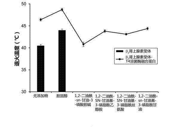
[0024] ①Take pure glycerol monooleate or a mixture of glycerol monooleate and one of the following lipid molecules out of the refrigerator at -20°C, and heat it to 37-40°C to form a liquid state; the aforementioned lipid molecules selected from 1,2-dioleoyl-sn-glycero-3-phosphocholine, 1,2-dioleoyl-SN-glycero-3-phosphatidylethanolamine, 1,2-dioleoyl-sn-glycerol Base-3-phosphatidylglycerol, 1,2-dioleoyl-SN-gl...

Embodiment 2

[0029] Example 2, the method described in Example 1, the mixture of glycerol monooleate and lipid molecules described in step (2) is prepared by the following method: the ratio of glycerol monooleate and the lipid molecules Evaporate most of the solvent in an appropriate amount of chloroform using nitrogen flow, then treat under vacuum at 21-23°C for at least 12 hours, and store at -20°C after removing the remaining residual chloroform.

Embodiment 3

[0030] Embodiment 3: experimental example.

[0031] 1. Expression and purification of adrenoceptor membrane protein

[0032] (1) Enter the "beta2 adrenergic receptor" search sequence on the NCBI website. According to the sequence published on the NCBI website, the full-length sequence of the gene encoding adrenoceptor in eukaryotic cells was obtained.

[0033](2) Transfer the target gene into the insect HEK293 cell expression system. HEK293 cells were cultured on plastic dishes at 37°C in Dulbecco's modified Eagle medium (Cellgro) with 5% carbon dioxide and 5% fetal bovine serum. After about 48 hours, add 1 microgram of vector pCDNA3 receptor plasmid and 3 microliters of Fugene6 reagent for transfection. After about 48 hours, the cells were washed with PBS, fixed with 4% paraformaldehyde, blocked with PBS + 2% goat serum, and permeabilized with PBS + 2% goat serum + 0.5% NONIDET P-40 (Sigma Company) .

[0034] (3) Technical advantage of modifying Eagle medium (Cellgro) ba...


PUM


Abstract

The invention relates to a method for measuring the stability of a membrane protein in a lipidic cubic sample through spectroscopy. The method comprises the following steps of: uniformly mixing a purified target protein into the lipidic cubic sample, and then carrying out a gradient heating/cooling process with temperature increased step by step on the lipidic cubic sample; cooling the lipidic cubic sample again and centrifugalizing the lipidic cubic sample at a high speed to ensure that water molecules enter the lipidic cubic sample again and the lipidic cubic sample is at a colorless and transparent state again when the lipidic cubic sample becomes turbid as water molecules are separated out of the lipidic cubic sample at every step of temperature rise; and then identifying the stability of the target protein by acquiring fluorescent signals of the lipidic cubic sample. The method disclosed by the invention is reasonable in design and high in operability, can realize that the stability of the protein can be directly measured in the lipidic cubic sample, and provides the data and theory support for subsequent crystal and correlational research; and a gene applied to the method disclosed by the invention can also provide the support for control research on various classified micromolecules.

Description

technical field [0001] The invention relates to the technical field of protein genetic engineering, in particular to a method for measuring the activity and stability of membrane proteins in lipid cube samples through spectroscopy. Background technique [0002] As early as 1960, researchers have proved that hydrophilic and hydrophobic amphiphilic molecules can form many different structures in water. In 1962, Luzzati's research group determined the structure of the layered phase and also obtained the electron microstructure of the hexagonal phase. Shortly thereafter, the structure of the lipid cubic phase was determined by X-ray techniques. In 1968, Luzzati and Canadian research experts conducted an X-ray study on the lipid-water system, and obtained a partial phase diagram of the lipid-water system as a function of lipid concentration and temperature. In 1980, Larsson proposed that the lipid bilayer membranes arranged in infinite cycles in cubic liquid crystals are very s...

Claims


Application Information

Patent Timeline
Patent Type & Authority Applications(China)
IPC IPC(8): G01N21/31G01N21/64G01N1/28
Inventor 刘伟许恒皓尹欣卢辰高嵩程斌
Owner 连云港脂立方生物医药研究所有限公司
Why Eureka
Try Eureka