

Rabbit hemorrhagic disease virus RT-PCR detection method
A technology of RT-PCR and rabbit hemorrhagic disease virus, which is applied in the direction of microorganism-based methods, biochemical equipment and methods, and microorganism measurement/inspection. Poor repeatability and other issues, to avoid false positive results, simple operation, easy to operate
- Summary
- Abstract
- Description
- Claims
- Application Information
AI Technical Summary
Problems solved by technology
Method used
Image

Examples
Embodiment 1
[0043] The preparation of embodiment 1 positive standard
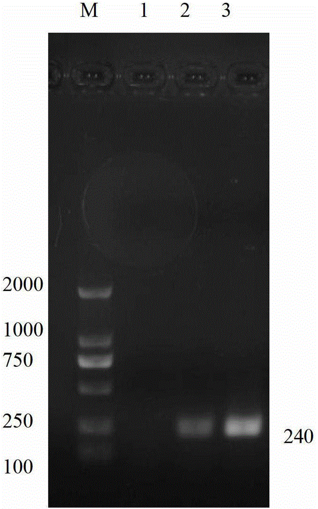
[0044] 1) Extract RNA from RHDV virus, and use primers with nucleotide sequences as shown in SEQ ID No.4 and 5 to perform one-step RT-PCR amplification to obtain a 240bp target fragment (such as figure 1 shown);
[0045] The PCR reaction system is:
[0046] Each 25 μL reaction liquid contains: 10×RT-PCR buffer 2.5 μL, ultrapure dNTP mixture (10 mM each) 1 μL, 5×RT-PCR enhancer 5 μL, 40U / μL RNase inhibitor 0.25 μL, 2.5U / μL Hotmaster DNA polymerase 1.25 μL, Quant reverse transcriptase 0.25 μL, 10 mmol / L upstream and downstream primers 0.5 μL each, RNA template 1 μL, RNase-free ddH 2 O supplemented to 25 μL;
[0047] The PCR reaction conditions are: 50°C, 30min, 94°C, 2min, 94°C, 1min, 51.7°C, 1min, 65°C, 1min, 35 cycles of amplification, 65°C, 10min;
[0048] 2) The target fragment was then cloned into the PGEM-T Easy vector (purchased from Promega), and transformed into the host cell Escherichia coli DH5α (purchased...
Embodiment 2
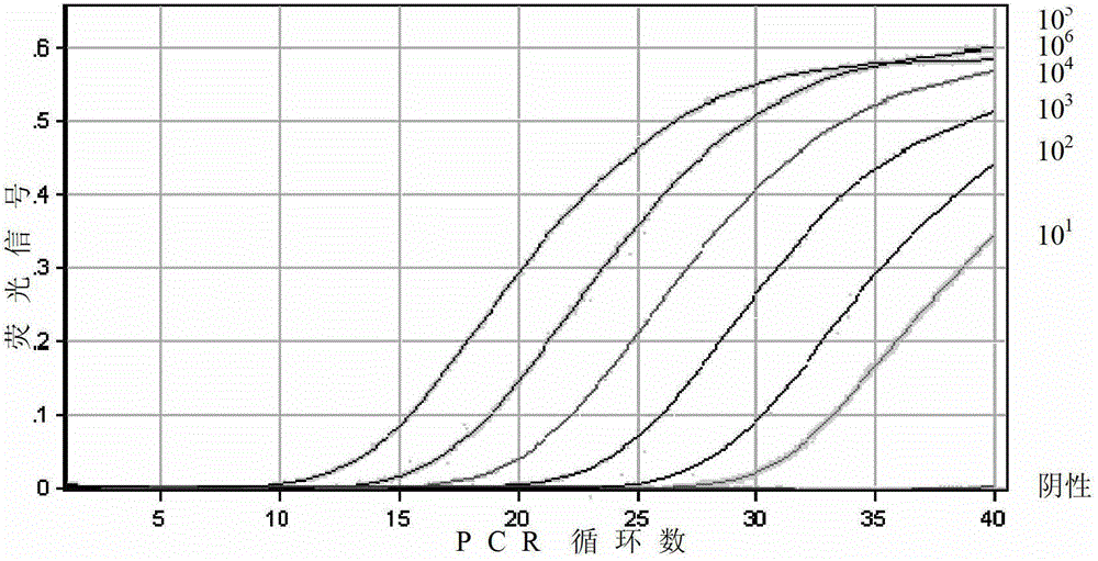
[0055] Example 2 standard curve drawing
[0056] The PCR primers were designed and screened according to GenBank (GenBank registration number: FJ794180) RHDV VP60 capsid protein gene sequence, and the specificity of each primer was confirmed. The nucleotide sequences of the primers are shown in SEQ ID No.1 and 2. Primers were synthesized by Shanghai Yingjun Biotechnology Co., Ltd. The length of the amplified fragment of the primer pair is 96bp.
[0057] Using the positive standard obtained in Example 1 as a template, using primers with nucleotide sequences as shown in SEQ ID No. 1 and 2 and fluorescent probe VP60p, a one-step RT-PCR method is used for detection. Test results such as image 3 shown. The reaction system in the above RT-PCR is:
[0058] Each 25 μL reaction solution contains: 2×Quant One Step Probe qRT-PCR Master Mix (Quant one-step fluorescent quantitative RT-PCR kit (probe method)) 12.5 μL, 2.5U / μL Hotmaster DNA polymerase 1 μL, 4U / μL Quant reverse transcri...
Embodiment 3
[0061] Embodiment 3 is to the detection of rabbit liver tissue sample
[0062] 1. Extraction of RNA from rabbit liver tissue samples
[0063] Take 0.2 mL of a 100-fold dilution of RHDV isolates (purchased from Jiangsu Academy of Agricultural Sciences), and inject intramuscularly into 3-month-old unimmunized rabbits. Three days later, some rabbits died. The liver tissues of 10 dead rabbits were collected, and the rabbit liver tissue samples were extracted using the RNAprep pure animal tissue total RNA extraction kit (purchased from Tiangen Biochemical Technology Co., Ltd.).
[0064] (1) Add 300 μL Lysis Buffer RL to 200 mg of liver tissue, thoroughly grind it into a homogenate, add 590 μL RNase-free ddH 2 O and 10 μL proteinase K, mix well and treat at 56°C for 10 min.
[0065] (2) Centrifuge the obtained liquid at 12000rpm for 5min, and take the supernatant.
[0066] (3) Slowly add 200 μL of absolute ethanol to the supernatant, mix well, put the obtained solution and the pr...
PUM
 Abstract
Description
Claims
Application Information