Method and kit for identifying sensitivity of antibody and clone cell strain

A sensitive, cell line technology, applied in chemical instruments and methods, specific peptides, biological testing, etc., can solve the problems of long operation time, limited economic affordability, unfavorable popularization, etc., achieve accurate results, reduce manpower and material resources. wasteful, simple effect

Inactive Publication Date: 2013-07-03
SHENZHEN MINDRAY BIO MEDICAL ELECTRONICS CO LTD +1
View PDF10 Cites 6 Cited by
  • Summary
  • Abstract
  • Description
  • Claims
  • Application Information

AI Technical Summary

Problems solved by technology

[0005] Immunofluorescence test includes direct immunofluorescence test and indirect immunofluorescence test, in which indirect immunofluorescence test needs to be completed with the help of secondary antibody. Long, cumbersome operation steps, and expensive experimental equipment, such as fluorescence microscope, flow cytometer
[0006] CN1660907A discloses three identification methods of monoclonal antibodies, including indirect immunofluorescence, dot immunoblotting and gold-labeled immunoelectron microscopy, in which both indirect immunofluorescence and gold-labeled immunoelectron microscopy require expensive equipment to cooperate with the judgment of the results. The dot immunoblotting method uses nitrocellulose membrane as a solid phase carrier for antigen-antibody reaction, and needs to obtain cell membrane protein lysate for adsorption. Since the protein in the cell membrane protein lysate is denatured, the identification result cannot reflect the monoclonal antibody and natural conformational protein sensitivity
[0007] CN1718588A discloses a method of monoclonal antibody for malignant tumor detection, which is realized by ELISA technology and immunoblotting technology, ELISA test technology uses recombinant protein as antigen for detection, and Western blotting test technology uses denatured protein as antigen Assays performed, neither test technique truly reflects the sensitivity of mAbs to native conformational proteins
[0008] CN101891806A discloses a screening method for the quality of monoclonal antibodies, which is realized by using indirect immunofluorescence technology. The judgment of indirect immunofluorescence test requires an expensive fluorescence microscope, and the economic affordability of general small clinical and scientific research institutions is limited. This method Not conducive to popularization
This method generally uses recombinant proteins as antigens, and the screened monoclonal antibodies are often not suitable for the binding reaction of natural conformational proteins, resulting in screening failure and waste of manpower and material resources
[0010] CN1693461A discloses the screening method of positive clonal cell strains in the preparation process of monoclonal antibody, is to use indirect enzyme-linked immunosorbent assay (ELISA) to carry out, this method generally is to use recombinant protein as antigen to screen, but because recombinant protein and natural conformation There are certain differences in proteins, so the positive clonal cell lines screened by ELISA cannot truly reflect the sensitivity of monoclonal antibodies and natural conformational proteins

Method used


Image

  • Method and kit for identifying sensitivity of antibody and clone cell strain
  • Method and kit for identifying sensitivity of antibody and clone cell strain
  • Method and kit for identifying sensitivity of antibody and clone cell strain

Examples


Embodiment

[0076] The present invention will be further described through specific examples below. The examples are only to illustrate the present invention, but not to limit the present invention in any sense.

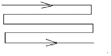
[0077] Unless otherwise stated, the reagent components used in the following examples are of analytical grade, the solvent used is deionized water, and the cell counting device used is a Nikon ANTI-MOULD inverted microscope. The schematic diagram of the cell counting method is as follows: figure 1 shown. The magnification of the cells under the microscope was 100×10. The format of the 96-well polystyrene plate described in the following examples is 400 uL / well.

Embodiment 1

[0079] The mouse anti-human CD3 monoclonal antibody to be tested, the well-identified mouse anti-human CD3 monoclonal antibody (positive control) and the negative control mouse IgG (basically not combined with CD3 antigen) were all coated with 10ug / plate for 96 Well polystyrene plate, set up two duplicate wells for each test, use carbonate buffer solution of pH 9.8 as the coating solution, place in a 37-degree incubator for 1 hour, take out the 96-well plate, and place it on a clean towel with 96 wells turned upside down After the liquid in the wells of the plate was patted dry, 100uL / well of the prepared lymphocyte suspension was added, and the concentration of the lymphocyte suspension was 6×10 7 / L, place on a shaker at room temperature and shake slightly for 30 minutes, use the cell washing solution to elute the reaction wells, add 200uL cell washing solution to each well, slightly blow the liquid in the well, then suck the liquid in the well, repeat Wash 3 times, use 4% n...

Embodiment 2

[0081] Coat the culture supernatant of the two mouse anti-human CD4 antibody cell lines to be tested and the negative control mouse IgG at 5ug / plate on a 96-well polystyrene plate, set up two duplicate wells for each test, and buffer with carbonate pH 9.0 As the coating solution, place it in a 37-degree incubator for 1.5 hours, take out the 96-well plate, place it on a clean towel and turn the 96-well plate upside down, pat dry the liquid in the well, add the prepared lymphocyte suspension 100uL / well, The concentration of lymphocyte suspension was 6×10 8 / L, place on a shaker at room temperature and shake slightly for 1 hour, use the cell washing solution to elute the reaction wells, add 200uL cell washing solution to each well, use a pipette with a 200uL range to slightly blow the liquid in the well, and then Aspirate the liquid in the well, repeat the washing 3 times, use 4% neutral formaldehyde fixative to fix the combined cells in the 96-well polystyrene plate, add 50uL of...


PUM

PropertyMeasurementUnit
strengthaaaaaaaaaa

Abstract

The invention discloses a method for identifying sensitivity of an antibody as well as quality of a clone cell strain in the preparation process of the antibody. The method comprises the following steps: acquiring a solid-phase carrier, cells and the antibody, adsorbing the antibody on the solid-phase carrier, incubating the cells and the antibody adsorbed on the solid-phase carrier, keeping cells that are combined with the antibody, dyeing and counting the cells combined with the antibody, and identifying the sensitivity of the antibody and the quality of the clone cell strain according to the cell counting result. The invention further provides a kit applicable to the method. The method intuitively reflects the level of combination of the antibody and a native conformation antigen, and the identifying method is simple, quick and intuitive.

Description

technical field [0001] The invention relates to the technical field of antibodies. More specifically, the present invention relates to a method for identifying antibody sensitivity, and a method for identifying positive clonal cell lines secreting target antibodies during antibody preparation. Background technique [0002] Antibodies are a type of globulin with immune function secreted by B cells that proliferate and differentiate into plasma cells. They exist in body fluids and mediate humoral immunity. Antibodies can specifically bind to corresponding antigens (such as pathogens), and exert immune effects with the participation of other immune molecules and cells. According to different properties, antibodies can be divided into monoclonal antibodies (hereinafter referred to as monoclonal antibodies) and polyclonal antibodies (hereinafter referred to as polyclonal antibodies). Monoclonal antibodies have the advantages of highly uniform physical and chemical properties, s...

Claims


Application Information

Patent Timeline
Patent Type & Authority Applications(China)
IPC IPC(8): G01N33/68
CPCG01N33/56972C07K16/00C07K16/2803C07K16/2809C07K16/2812C07K16/289G01N33/554G01N33/6854G01N2333/7051G01N2333/70514G01N2333/70589G01N2333/70596
Inventor 赵玉梅董丽芳雷霆
Owner SHENZHEN MINDRAY BIO MEDICAL ELECTRONICS CO LTD
Why Eureka
Try Eureka