Micro-fluidic chip for cell culture as well as preparation method and application of micro-fluidic chip

A microfluidic chip, cell culture technology, applied in tissue cell/virus culture devices, biochemical equipment and methods, tissue culture, etc. Easy-to-clean, simple to make, low-cost effects

Inactive Publication Date: 2014-12-17
CHONGQING UNIV
View PDF2 Cites 0 Cited by
  • Summary
  • Abstract
  • Description
  • Claims
  • Application Information

AI Technical Summary

Problems solved by technology

[0006] At present, most of the microfluidic chips are processed by soft lithography, and the production process is relatively complicated and needs to be completed by some expensive and high-precision instruments.
For example: the production of its mask needs to be completed with the help of a high-precision film printer; the process of making a positive mold using SU-8 negative photoresist lithography is quite complicated and cumbersome; most chips must be bonded with the help of a plasma surface processor. Complete packaging; chip filling must be completed with the help of precision instruments, etc.
Moreover, such chips are difficult to clean and can only be used once, which increases the cost of the experiment to a certain extent.
All of these have limited the application of microfluidic chips in ordinary experiments.
In addition, since the packaging needs to be completed by bonding with a plasma surface processor, a thicker glass slide must be used as the bottom substrate, which is not conducive to laser confocal observation

Method used


Image

  • Micro-fluidic chip for cell culture as well as preparation method and application of micro-fluidic chip
  • Micro-fluidic chip for cell culture as well as preparation method and application of micro-fluidic chip
  • Micro-fluidic chip for cell culture as well as preparation method and application of micro-fluidic chip

Examples


Embodiment 1

[0044] Example 1 Structure of a cell culture microfluidic chip

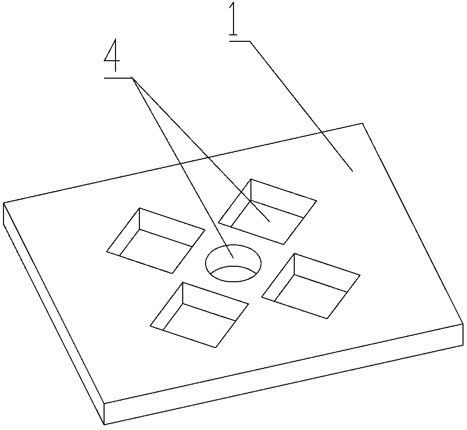
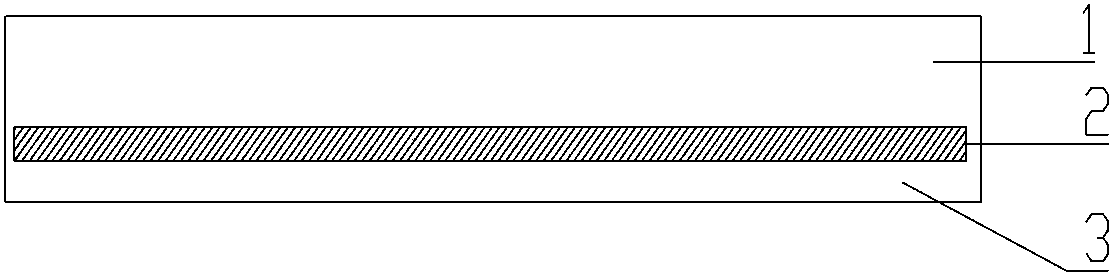
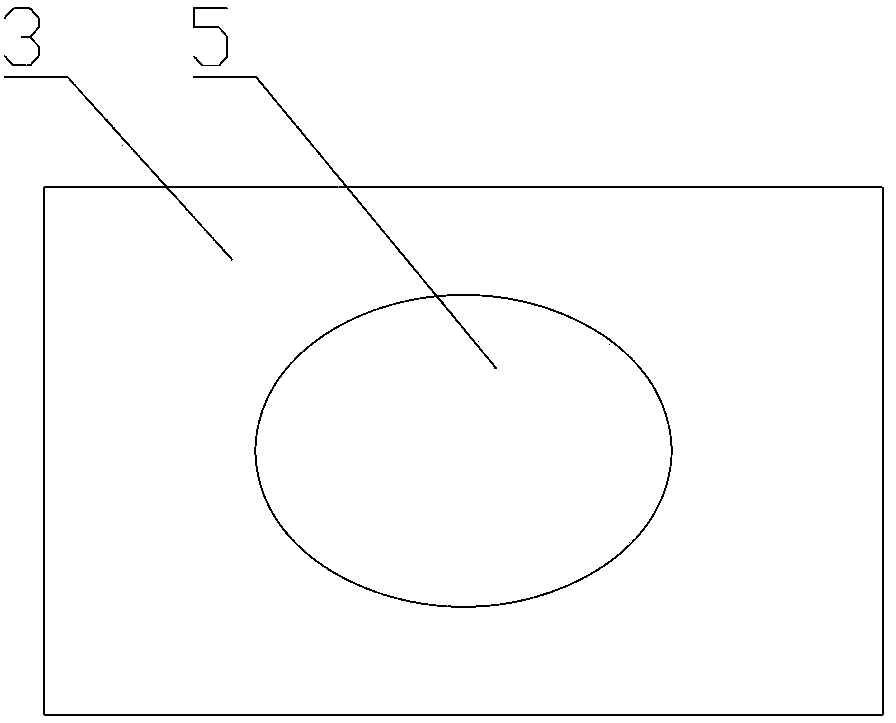
[0045] Microfluidic chip for cell culture, including cell culture layer 1, base layer 2 and clamping layer 3 ( figure 2 shown), the three sides between the cell culture layer 1 and the clamping layer 3 are sealed into a jacket structure, the base layer 2 is set in the jacket structure in a removable manner, and the base layer and the cell culture layer Sealing, 5 holes are distributed on the cell culture layer, 1 hole in the middle, and 4 holes are distributed around it, each hole and the base layer jointly form a cell culture chamber 4 ( figure 1 shown). In addition, the clamping layer is provided with an observation hole 5, which is convenient for visual observation under a microscope ( image 3 shown).

[0046] The microfluidic chip is made of PDMS material, 3cm in length, 2cm in width, and 0.5cm in height, and is equipped with 5 cell culture chambers, and each cell culture chamber is penetrated by filling...

Embodiment 4

[0064] Two-dimensional culture of embodiment 4 human umbilical vein endothelial cells

[0065] Pour collagen or matrigel into the cell culture chamber 4, and after gel formation, directly add the human umbilical vein endothelial cell line suspension into the cell culture chamber 4, use DMEM complete medium for culture, and conduct two-dimensional culture, observe cell shape, such as Image 6 shown.

Embodiment 5

[0066] Example 5 Three-dimensional culture of human umbilical vein endothelial cells

[0067] Mix human umbilical vein endothelial cells with collagen or Matrigel and pour them into the middle cell culture chamber 4. After the gel is formed, add DMEM complete medium to the middle cell culture chamber 4 for culture and observe the cells form and behavior, such as Figure 7 shown.


PUM


Abstract

The invention relates to the field of biochips and in particular relates to a micro-fluidic chip for cell culture as well as a preparation method and application of the cell culture. The micro-fluidic chip comprises a cell culture layer (1), a substrate layer (2) and a clamping layer (3); materials required for preparation of the micro-fluidic chip are conventional slides in a laboratory and PDMS (Polydimethylsiloxane); and the micro-fluidic chip is prepared by using the five steps of arranging the slides, preparing a PDMS preforming solution, molding, forming and packaging. The micro-fluidic chip is simple to manufacture and low in manufacturing cost; and the micro-fluidic chip can be manufactured without the assistance of expensive instruments. The microfluidic chip provided by the invention can be used for realizing multiple modes of cell culture, accurately controlling the parameters of cells in a microenvironment by a smart configuration design and a microfluidic control technology and investigating the influence of the cell microenvironment on cell behaviors.

Description

technical field [0001] The invention relates to a microfluidic chip, in particular to a cell culture microfluidic chip for cell microenvironment research, as well as its preparation method and application in cell microenvironment research. Background technique [0002] The cellular microenvironment is a complex collection of multiple factors, which play an important role in the behavior and function of cell proliferation, differentiation, migration and metabolism. These behaviors of cells are closely related to important physiological and pathological processes such as embryonic development, angiogenesis, tissue repair, immune defense, and tumorigenesis. In recent years, the study of cell microenvironment has become a research hotspot and difficulty. The microenvironment in which cells live is very complex, and its complexity can be summarized as: the influence of the three-dimensional micro-scale space in which cells live on cell behavior; cell interaction between differen...

Claims


Application Information

Patent Timeline
Patent Type & Authority Patents(China)
IPC IPC(8): C12M3/00C12N5/00
Inventor 陈斯佳蔡绍皙张利光邹米莎赵毅何腾龙陈龙聪李博柯明
Owner CHONGQING UNIV
Why Eureka
Try Eureka