Magneto-optical bimodal imaging probe rare earth nanoparticle, and preparation method and application thereof

A magneto-optical dual-mode and nanoparticle technology, which is applied in nuclear magnetic resonance/magnetic resonance imaging contrast agents, luminescent materials, fluorescence/phosphorescence, etc., can solve the problems of degradation of organic fluorescent molecules, inability to meet long-term tracers, cumbersome preparation methods, etc. problems, to achieve the effects of low toxicity and side effects, good clinical application prospects, and high quantum yield

Active Publication Date: 2013-09-04
NANJING UNIV
View PDF5 Cites 9 Cited by
  • Summary
  • Abstract
  • Description
  • Claims
  • Application Information

AI Technical Summary

Problems solved by technology

Although the nanoparticles prepared by the above method can achieve the effect of photomagnetic imaging, the preparation method is cumbersome, and the organic fluorescent molecules may be degraded and quenched during the in vivo imaging process, which cannot meet the requirements of long-term tracer

Method used


Image

  • Magneto-optical bimodal imaging probe rare earth nanoparticle, and preparation method and application thereof
  • Magneto-optical bimodal imaging probe rare earth nanoparticle, and preparation method and application thereof
  • Magneto-optical bimodal imaging probe rare earth nanoparticle, and preparation method and application thereof

Examples


Embodiment 1

[0021] Embodiment 1. Preparation of rare earth nanoparticles of a magneto-optic dual-mode imaging probe:

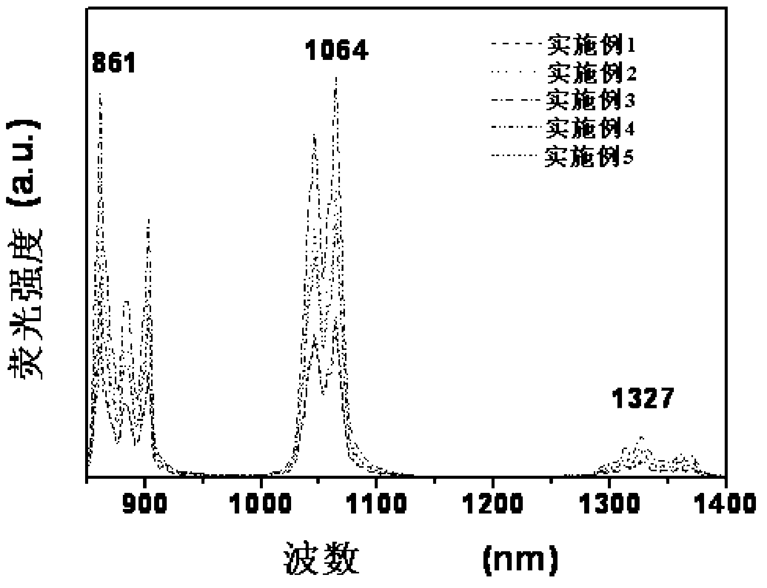
[0022] 1.78mmol of La(NO 3 ) 3 ·6H 2 O, 1.19 mmol of Gd(NO 3 ) 3 ·6H 2 O and 0.03mmol Nd(NO 3 ) 3 ·6H 2 O was dissolved in 10 mL of distilled water and mixed with 20 mL of ethanol. Then, the mixture of 10mL NaF aqueous solution (1M) and 5mL10wt% polyethyleneimine aqueous solution was added dropwise to the above solution, and the obtained mixed solution was stirred for 10min and then transferred to a 50ml reactor, sealed and heated to 180°C for 10h. Finally, the reactor was cooled to room temperature. The precipitate was separated by centrifugation and washed several times with ethanol and water. Finally, vacuum-dried to obtain polyethyleneimine-coated La 0.6 Gd 0.4 f 3 : Nd1% rare earth nanoparticles, (the element content of the product is measured by inductively coupled plasma mass spectrometry, the same below), the basic physical and chemical parameters are...

Embodiment 2

[0023] Example 2. Preparation of a magneto-optical dual-mode imaging probe rare earth nanoparticles:

[0024] 0.56mmol of La(NO 3 ) 3 ·6H 2 O, 2.24 mmol of Gd(NO 3 ) 3 ·6H 2 O and 0.2mmol Nd(NO 3 ) 3 ·6H 2 O was dissolved in 10 mL of distilled water and mixed with 20 mL of ethanol. Then, 10mL of NaF aqueous solution (1M) and 5mL of 10wt% polyvinyl alcohol aqueous solution were mixed and added dropwise to the above solution, and the obtained mixed solution was stirred for 10 minutes and then transferred to a 50ml reaction kettle, sealed and heated to 150°C for 4 hours. Finally, the reactor was cooled to room temperature. The precipitate was separated by centrifugation and washed several times with ethanol and water. Finally, the polyvinyl alcohol-coated La was obtained by vacuum drying. 0.2 Gd 0.8 f 3 : Nd7% rare earth nanoparticles, the basic physical and chemical parameters are shown in Table 1.

Embodiment 3

[0025] Example 3. Preparation of a magneto-optical dual-mode imaging probe rare earth nanoparticles:

[0026] 0.81mmol of La(NO 3 ) 3 ·6H 2 O, 1.89 mmol of Gd(NO 3 ) 3 ·6H 2 O and 0.3mmolNd(NO 3 ) 3 ·6H 2 O was dissolved in 10 mL of distilled water and mixed with 20 mL of ethanol. Then, the chitosan aqueous solution mixture of 10mL NaF aqueous solution (1M) and 5mL10wt% was added dropwise to the above-mentioned solution, and the obtained mixture was stirred for 10min, then transferred to a 50ml reactor, sealed and heated to 250°C for 12h. Finally, the reactor was cooled to room temperature. The precipitate was separated by centrifugation and washed several times with ethanol and water. Finally, vacuum drying yielded chitosan-coated La 0.3 Gd 0.7 f 3 : Nd10%, see Table 1 for basic physical and chemical parameters.


PUM

PropertyMeasurementUnit
radiusaaaaaaaaaa
thicknessaaaaaaaaaa
particle diameteraaaaaaaaaa

Abstract

The invention relates to a rare earth nanoparticle with multimodal imaging functions of magnetic resonance imaging (MRI) and near infrared refraction (NIR) imaging, and a preparation method and the application thereof. The method is characterized in that macromolecules, namely synthetic polymers or biomacromolecules are used as a protecting agent; fluoride LaxGd1-xF3 is used as a matrix material, wherein x ranges from 0.1 to 0.9; neodymium ions, namely Nd3 <+> are used as a luminescent centre; and the rare earth nanoparticle is a water-soluble rare earth nanoparticle with the particle size of 10-100 nm. A fluorescence spectrum of the doped rare earth nanoparticle LaF3 / GdF3 : Nd has two very strong characteristic peaks (863 nm and 892 nm as well as1042 nm and 1057 nm) when being displayed in a near-infrared region with wavelength of 700-1200 nm, in-vitro magnetic resonance imaging data show that the rare earth nanoparticle has higher longitudinal relaxation rate, so that the rare earth nanoparticle can be used for preparing a near-infrared and magnetic resonance bimodal contrast agent for a living body.

Description

technical field [0001] The invention relates to the field of magneto-optical dual-mode imaging probes, in particular to a fluoride La x Gd 1-x f 3 (x=0.1~0.9) is the matrix material, neodymium ions (Nd 3+ ) as the rare earth-doped nanoparticle LaF as the luminescent center 3 / GdF 3 : Nd and its preparation method, and the application of the nanoparticle as a living near-infrared and magnetic resonance dual-mode contrast agent. Background technique [0002] Cancer is one of the major diseases threatening human health at present. Surgery is still the preferred treatment method, and resection of the primary tumor is a common operation. However, a large number of clinicopathological evidences show that there are quite a few early cancers where no obvious lesion area can be found. Moreover, the tumor often grows in an infiltrating manner, resulting in blurred boundaries with normal tissues, which brings great difficulties to surgical resection. Incomplete tumor resection ...

Claims


Application Information

Patent Timeline
Patent Type & Authority Applications(China)
IPC IPC(8): C09K11/85C09K11/02A61K49/06G01N21/64
Inventor 丁寅陈洪渊
Owner NANJING UNIV
Why Eureka
Try Eureka