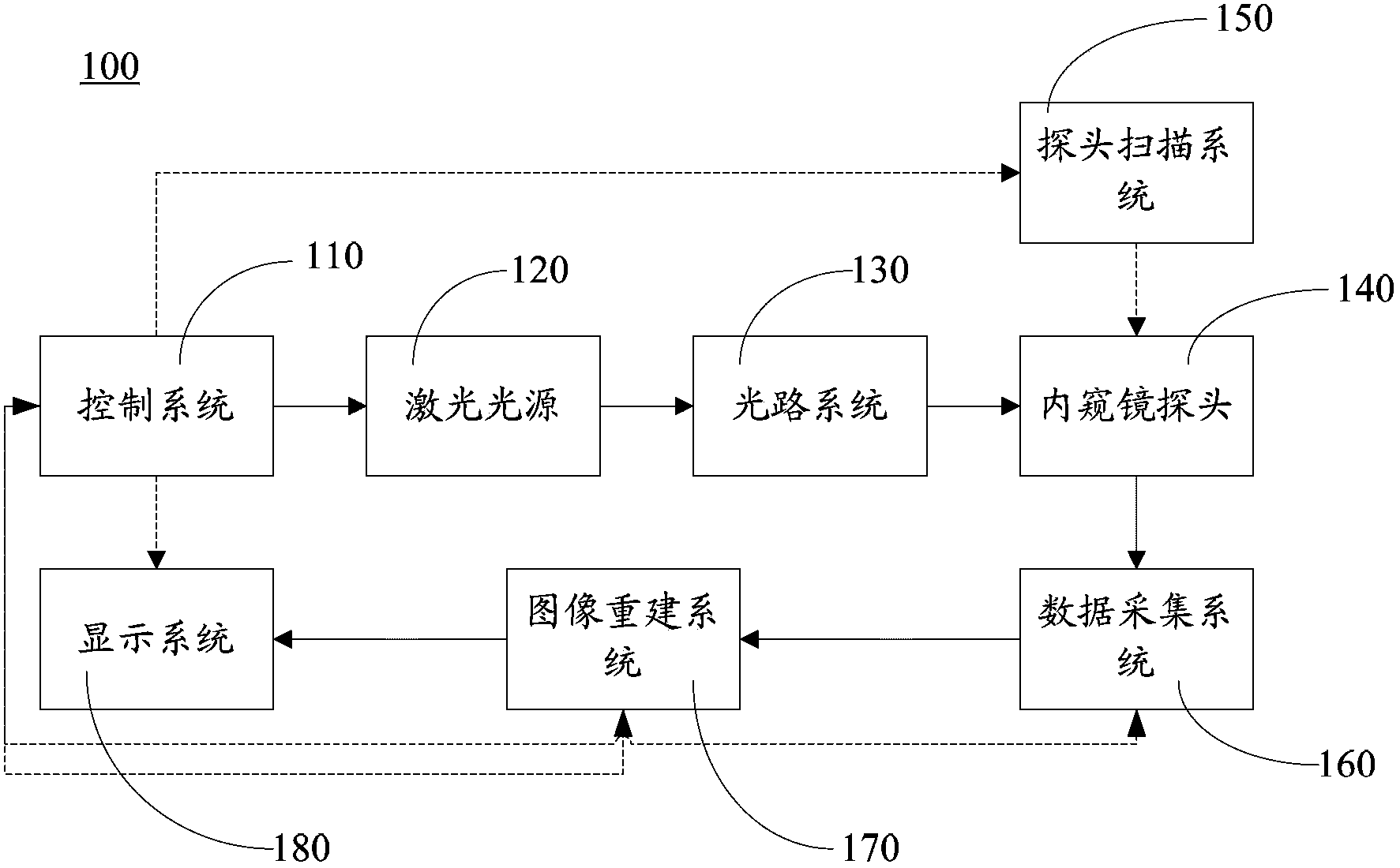
Photoacoustic endoscope
An endoscope and laser technology, used in catheters, operations, etc., can solve the problems of low imaging resolution, low soft tissue contrast, and can not fully meet clinical needs, and achieve the effect of high image resolution and good optical resolution.
- Summary
- Abstract
- Description
- Claims
- Application Information
AI Technical Summary
Problems solved by technology
Method used
Image

Examples
Embodiment 1
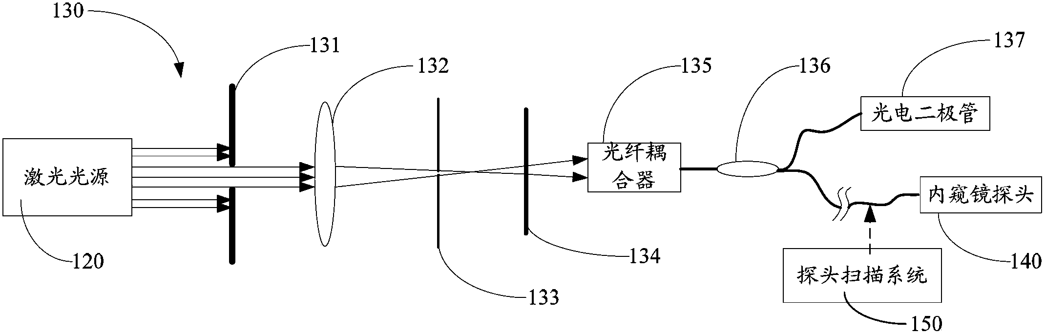
[0041] Such as image 3 and Figure 4 As shown, an optical fiber 212 is disposed within a fiber optic guide 214 . Further, in this embodiment, the fiber optic guide 214 is provided with a flexible guide 216 . The optical fibers are wrapped in a flexible conduit 216 .
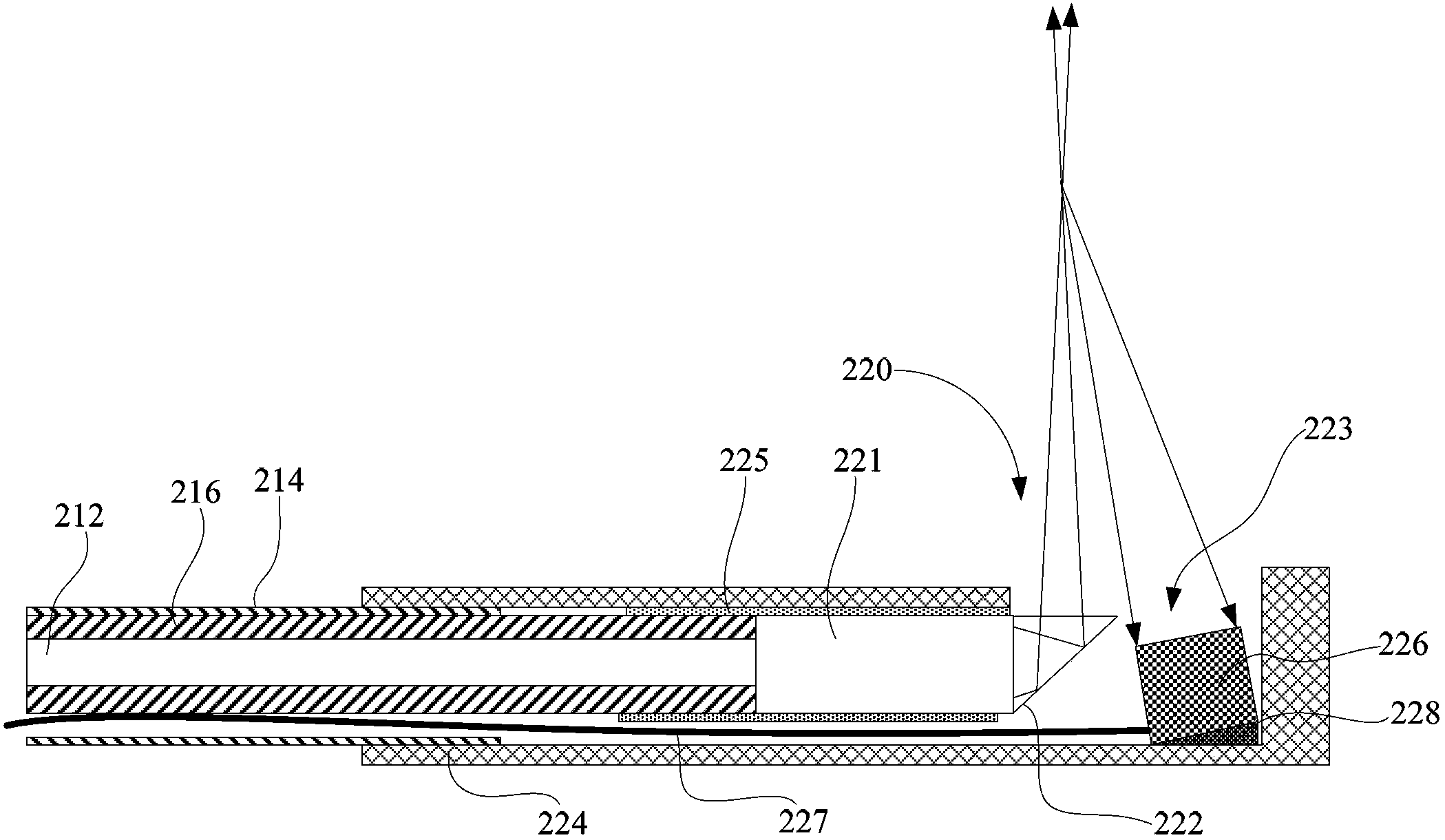
[0042] The endoscopic probe 220 includes an optical component and a fixing component (not shown in the figure). Wherein, the optical components include a self-focusing lens 221 and a reflector 222 . The fixing assembly includes a rigid conduit 224 with a photoacoustic window 223 at its free end and a plastic sleeve 225 inside the rigid conduit 224 .
[0043] Fiber optic conduit 214 is partially inserted into rigid conduit 224 . The outer wall of the fiber optic guide 214 inserted into the rigid guide 224 is socketed with the inner wall of the rigid guide 224 . The ends of the flexible conduit 216 and the optical fiber 212 are flush, and pass through the optical fiber conduit 214 , partly inserted into the ...
Embodiment 2
[0048] Such as Figure 5 As shown, the structure of the endoscopic probe and optical fiber of the present embodiment is similar to that of Embodiment 1, and the difference is that the rectangular prism 422 in the present embodiment is different from the reflector 222 of the isosceles rectangular prism used in Embodiment 1, Therefore, in this embodiment, without the adjustment member 228, it can be ensured that the ultrasonic transducer 426 can vertically receive the excited ultrasonic signal of the target tissue.
Embodiment 3
[0050] Such as Figure 6 As shown, an optical fiber 512 is disposed within a fiber optic guide 514 . Further, in this embodiment, a flexible conduit 516 is provided in the fiber optic conduit 514 . The optical fibers are wrapped in a flexible conduit 516 .
[0051] The endoscopic probe 520 includes an optical component and a fixing component (not shown in the figure). Wherein, the optical components include a self-focusing lens 521 and a reflector 522 . The fixing assembly includes a rigid conduit 524 with a photoacoustic window 523 at its free end and a plastic sleeve 525 inside the rigid conduit 524 .
[0052] The ends of the flexible conduit 516 and the optical fiber 512 are flush, and pass through the optical fiber conduit 514 , partly inserted into the plastic sleeve 525 , and coaxially connected with the self-focusing lens 521 at the end. The self-focusing lens 521 is wrapped in a plastic sleeve 525 and coaxially connected with the flexible conduit 516 and the optica...
PUM
| Property | Measurement | Unit |
|---|---|---|
| Wavelength | aaaaa | aaaaa |
Abstract
Description
Claims
Application Information
 - Generate Ideas
- Intellectual Property
- Life Sciences
- Materials
- Tech Scout
- Unparalleled Data Quality
- Higher Quality Content
- 60% Fewer Hallucinations
Browse by: Latest US Patents, China's latest patents, Technical Efficacy Thesaurus, Application Domain, Technology Topic, Popular Technical Reports.
© 2025 PatSnap. All rights reserved.Legal|Privacy policy|Modern Slavery Act Transparency Statement|Sitemap|About US| Contact US: help@patsnap.com