
Method for detecting DNA (deoxyribonucleic acid) through high sensitivity Raman spectrum

A sensitive Raman and spectral detection technology, applied in the field of high-sensitive Raman spectral detection of DNA, can solve the problems of light quenching, limit the application of fluorescence analysis technology, etc., and achieve the effects of simple operation, improved detection sensitivity, and low price.

Inactive Publication Date: 2014-01-22
NANJING UNIV
View PDF4 Cites 9 Cited by
  • Summary
  • Abstract
  • Description
  • Claims
  • Application Information

AI Technical Summary

Problems solved by technology

However, its own light quenching, complex operation and spectral overlap of different signals limit the application of fluorescence analysis technology

Method used


Image

  • Method for detecting DNA (deoxyribonucleic acid) through high sensitivity Raman spectrum

Examples


Embodiment 1

[0023] Embodiment 1: the preparation of the Raman dye-silver nanocomposite of primer modification

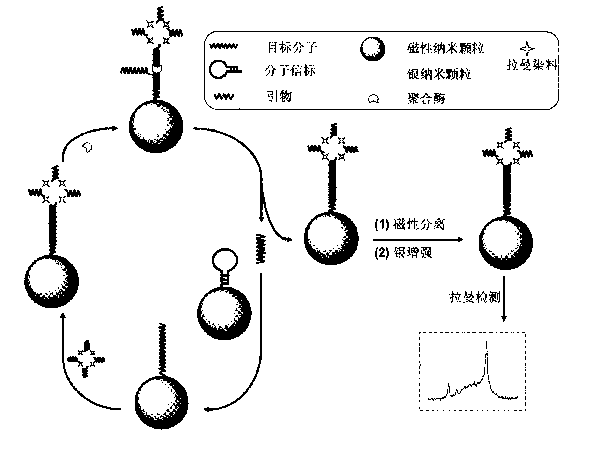
[0024] After boiling 200mL of 1.0mM silver nitrate solution, 5mL of 35mM sodium citrate solution was added under stirring, and the mixture was stirred and heated for 1 hour to obtain a colloidal solution of silver nanoparticles, which was stored in the dark at 4°C. Further, 100 μL of sulfhydryl-modified primers (10 μM) were added to 1 mL of colloidal solution of silver nanoparticles, stirred for 18 hours, and purified by centrifugation at 8000 rpm for 15 minutes for three times, and the obtained primer-modified silver nanoparticles were redispersed in 1 mL 10mM PBS solution. Then, 4 μL of 1 mM 4-mercaptobenzoic acid solution was added to this solution, stirred for 12 hours, and centrifuged and purified three times to obtain the primer-modified Raman dye-silver nanocomposite, which was re-introduced into 1 mL of 10 mM PBS solution for use.

Embodiment 2

[0025] Example 2: Immobilization of molecular beacons on the surface of magnetic beads

[0026] Disperse 50 μL of carboxylated magnetic nanoparticles into 1 mL of PBS (0.01M, pH 7.4), dropwise into 200 μL of 0.1M 1-ethyl-3-(3-dimethylaminopropyl)-carbodiimide hydrochloride Saline solution, 400 μL 0.05M N-hydroxysuccinimide solution and 150 μL 5.4 μM molecular beacon, stirred at room temperature for 4 hours, separated magnetic nanoparticles using a magnetic field, and washed several times with water to obtain molecular beacon-modified magnetic nanoparticles , redispersed in 1 mL of PBS (0.01M, pH 7.4) and stored at 4°C until use.

Embodiment 3

[0027] Embodiment 3: Raman spectroscopy detects DNA

[0028] (1) In 10 μL molecular beacon-modified magnetic nanoparticle dispersion, 10 μL DNA solution of different concentrations, 5 μL primer-modified Raman dye-silver nanocomposite solution, 2 μL polymerase (4 U) and dNTPs (each Nucleotides are all 12 μM), incubated at 37°C for 100 minutes, and then separated by a magnetic field to replace the strands immobilized on the surface of the magnetic nanoparticles;

[0029] (2) The chain substitution product was mixed with 10 μL of the mixture containing 1:1 silver enhancement solutions A and B, and Raman detection was performed after 2 minutes of silver enhancement reaction.

[0030] (3) Record the Raman signals of different concentrations of DNA solutions and samples, obtain the working curve of DNA detection, and calculate the concentration of DNA in the sample.

[0031] (4) By comparing the Raman signals at the same DNA concentration, the single base mismatch or unpaired DNA s...


PUM


Abstract

The invention relates to a method for detecting DNA (deoxyribonucleic acid) through high sensitivity Raman spectrum. According to the method, chains on the surfaces of magnetic beads are used for substituting the potentiation of polymerization and silver so as to realize dual amplification of the signal. The magnetic beads are used for fixing molecular beacons and separating chain substituted products. After hybridization of the target DNA and the molecular beacons, beacon rings are opened, raman dye-silver nano-composite modified by a primer is connected with the magnetic beads through hybridization of the primer and the stem end of the beacon so as to induce the DNA chain to carry out polymerization reaction to release target molecules, and the released DNA molecules and the molecular beacon of the other magnetic bead are hybridized so as to generate the other polymerization circulation. After the circulation chain substitutes polymerization, a separation chain substitutes the product, a silver nanoparticle shell is deposited on the surface of the nano-composite, so as to enhance the raman signal of the dye. The correlation between the raman signal and the concentration and sequence of the target DNA are used for DNA analysis. The concentration range of DNA detected by the method is 10-13-10-8mol L-1, and the method can be used for distinguishing completely matched target DNA and single-base mismatch DNA, and has certain application value.

Description

1. Technical field [0001] The invention is a method for detecting DNA with high-sensitivity Raman spectrum. It performs dual signal amplification through chain substitution polymerization on the surface of magnetic beads and silver enhancement, uses an external magnetic field to separate chain substitution products on magnetic beads, and uses Raman spectroscopy for highly sensitive DNA detection. 2. Background technology [0002] DNA is the bearer of genetic information, and the variation of base sequences in DNA molecules is related to many genetic diseases of human beings. Therefore, analyzing the DNA of a specific sequence and detecting the base mutation has far-reaching significance in gene screening, early diagnosis of genetic diseases, and the like. DNA hybridization is an important method for nucleic acid concentration detection and structural analysis. DNA hybridization detection techniques include fluorescence, chemiluminescence, electrochemistry, and surface plas...

Claims


Application Information

Patent Timeline
IPC IPC(8): G01N21/65
Inventor 鞠熀先高丰雷朱珠雷建平
Owner NANJING UNIV
Learn More