Method for detecting activation peroid markers of T lymphocyte in human peripheral blood

A lymphocyte and human peripheral blood technology, applied in the field of medical testing, can solve the problems that the test results cannot be directly compared, the absolute number of antigens or molecules cannot be expressed, and the qualitative analysis of fluorescence intensity cannot accurately reflect the characteristics of dynamic changes, etc.

Inactive Publication Date: 2014-02-26
AFFILIATED HUSN HOSPITAL OF FUDAN UNIV
View PDF1 Cites 7 Cited by
  • Summary
  • Abstract
  • Description
  • Claims
  • Application Information

AI Technical Summary

Problems solved by technology

However, the above results fail to express the absolute amount of a certain antigen or molecule on cells or particles
In addition, the expression of T lymphocyte activation antigens CD69, CD25, and CD71 is a continuous process of change, and the qualitative analysis of fluorescence intensity cannot accurately reflect its dynamic change characteristics, and the test results between various laboratories cannot be directly compared

Method used


Image

  • Method for detecting activation peroid markers of T lymphocyte in human peripheral blood
  • Method for detecting activation peroid markers of T lymphocyte in human peripheral blood
  • Method for detecting activation peroid markers of T lymphocyte in human peripheral blood

Examples


Embodiment 1

[0027] Example 1 The expression levels of CD69, CD25 and CD71 in healthy people

[0028] ①Sample collection: 5 healthy volunteers were selected, 3ml of peripheral venous blood was drawn, and placed in K 2 EDTA anticoagulant tube;

[0029] ②Take 100 μl anticoagulant blood, add 2ml RPMI1640 cell culture medium and 20μl ConA stimulator, 37°C, 5% CO 2 Cultivate in the incubator for 24h;

[0030] ③ Add 2 μl each of antihuman-CD3-PerCP-5.5, antihuman-CD69PE, antihuman-CD25PE and antihuman-CD71PE, incubate in the dark for 30 minutes, then add 3ml of erythrocyte lysate, and let stand in the dark for 10 minutes

[0031] ④ Centrifuge at 477g for 10min, discard the supernatant;

[0032] ⑤Add 2ml of PBS to wash, centrifuge at 477g for 10min, discard the supernatant, and repeat this process twice;

[0033] ⑥Add 100μl PBS to resuspend the cells, to be tested;

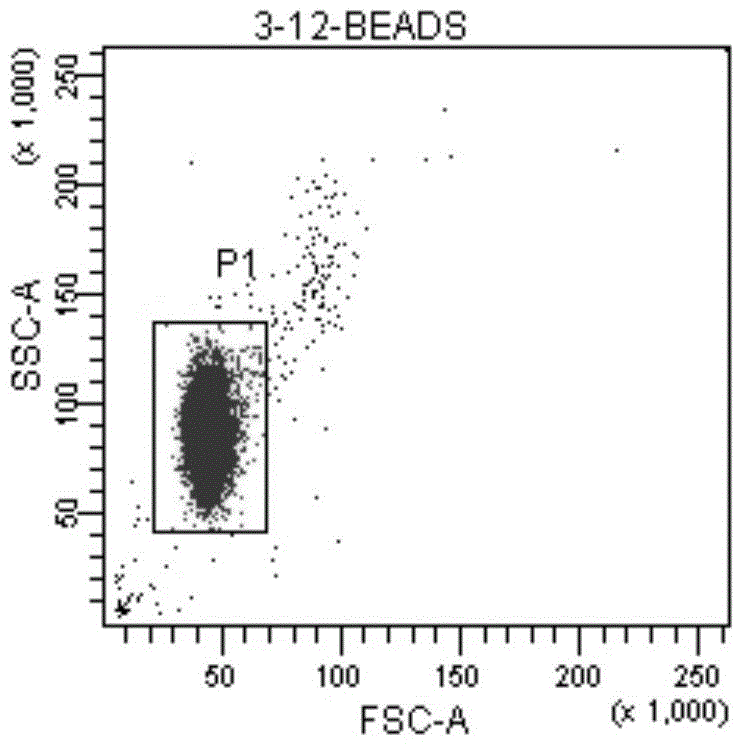
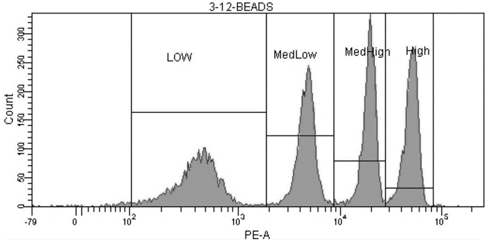
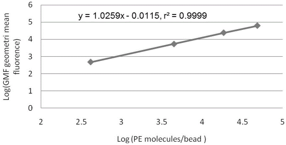
[0034] ⑦Preparation of standard microsphere solution: add 1ml of PBS to the measurement tube containing QuantiBRITE PE microsp...

Embodiment 2

[0040] Example 2 Expression of CD69, CD25 and CD71 in peripheral blood of kidney transplant patients

[0041] ①Sample collection: 5 kidney transplant patients were selected, 3ml of peripheral venous blood was drawn, and placed in K 2 EDTA anticoagulant tube;

[0042] ②Take 100μl anticoagulant blood, add 2ml RPMI1640 medium and 20μl ConA stimulator, 37℃, 5%CO 2 Cultivate in the incubator for 24h;

[0043]③ Add 2 μl each of antihuman-CD3-PerCP-5.5, antihuman-CD69PE, antihuman-CD25PE and antihuman-CD71PE antibodies, incubate in the dark for 30 minutes, then add 3ml of erythrocyte lysate, and let stand in the dark for 10 minutes

[0044] ④ Centrifuge at 477g for 10min, discard the supernatant;

[0045] ⑤Add 2ml of PBS to wash, centrifuge at 477g for 10min, discard the supernatant, and repeat this process twice;

[0046] ⑥Add 100μl PBS to resuspend the cells, to be tested;

[0047] ⑦Preparation of standard microsphere solution: add 1ml of PBS to the measurement tube containing...


PUM


Abstract

The invention belongs to the technical field of medical tests, and relates to a method for simultaneously determining activation markers CD69, CD25 and CD71 of T lymphocyte in human peripheral blood by employing quantitative flow cytometry. The method comprises the following steps: firstly, cultivating and stimulating peripheral blood; then carrying out dyeing treatment on the cultivated and stimulated peripheral blood, and carrying out red blood cell cracking and washing on the dyed sample; and finally carrying out quantitative flow detection on the washed sample. Meanwhile, a standard curve is built according to QuantiBRITE PE microspheres, and specific expression quantity of antigens on each cell surface is calculated through the standard curve. By adopting the method disclosed by the invention, the antigen expression quantity of the CD69, CD25 and CD71 at the surface of the human T lymphocyte can be accurately and sensitively determined, and immunological characteristics of the detected T cells can be reflected. Thus, certain immunological basis and detection index are supplied for clinical research.

Description

technical field [0001] The invention belongs to the technical field of medical detection, and in particular relates to a method for detecting T lymphocyte activation markers in human peripheral blood. Especially the method for simultaneously detecting T lymphocyte activation markers CD69, CD25 and CD71 in human peripheral blood by quantitative flow cytometry. Background technique [0002] Studies have shown that the safety range (ie, therapeutic window) of immunosuppressants is narrow, and pharmacokinetic parameters such as blood drug concentration vary greatly between individuals and within individuals, and serious adverse reactions such as infection or rejection are prone to occur, requiring therapeutic drug monitoring ( Therapeutic Drug Monitoring, TDM) and individualized drug delivery. At present, the therapeutic drug monitoring of immunosuppressants is usually carried out by blood concentration monitoring. However, studies in recent years have found that the same bloo...

Claims


Application Information

Patent Timeline
Patent Type & Authority Applications(China)
IPC IPC(8): G01N33/533G01N21/64
CPCG01N33/9493
Inventor 焦正范琳琳邱晓燕钟明康张明
Owner AFFILIATED HUSN HOSPITAL OF FUDAN UNIV
Why Eureka
Try Eureka