

Larva paraffin section forming method
A technology for paraffin sectioning and larvae, applied in the field of microscopic tissue sectioning, can solve the problems of difficulty in observing the complete spatial structure of worms, easy deformation or fragmentation of the digestive tract, limited tissue differentiation, etc., and achieves good transparent processing effect and tissue differentiation High-precision, short-time-consuming effect
- Summary
- Abstract
- Description
- Claims
- Application Information
AI Technical Summary
Problems solved by technology
Method used
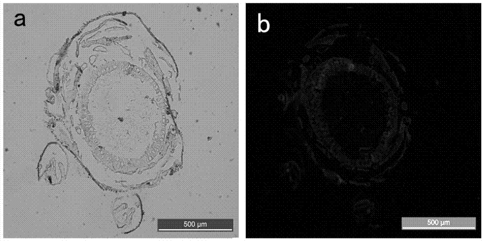
Image

Examples
Embodiment 1
[0060] In the present embodiment, the paraffin section method of insect larvae comprises the following steps:
[0061] 1. Tissue fixation
[0062] Prepare a 4% paraformaldehyde solution in a chemical fume hood as a tissue fixative, and the fixative is prepared for immediate use. The preparation process of the tissue fixative is: in 100 ml of PBS buffer (PBS buffer: NaCl 137mmol / L, KCl 2.7 mmol / L, Na 2 HPO 4 10mmol / L, KH 2 PO 42mmol / L, pH value 5~7), add solid sodium hydroxide particles to make the pH value reach 11, then heat to 70°C in a microwave oven, add 4g of paraformaldehyde particles, shake quickly until dissolved, and use an ice water bath Cool the solution, adjust the pH value to 7 with concentrated sulfuric acid, finally add 30 μl Tween20, and cool the prepared tissue fixative solution to 4°C;
[0063] The fixed procedure is as follows: Put the tested worms (3rd instar larvae of cotton bollworm indoors, breeding room temperature 27°C, relative humidity 60%, lig...
PUM
 Abstract
Description
Claims
Application Information