Improved antibody chip kit capable of quantitatively detecting multiple cell factors synchronously

A cytokine and antibody chip technology, applied in the field of biomedicine, can solve the problems of less specimen consumption, cumbersome operation, and low sensitivity, and achieve the effects of reducing background interference, increasing stability, and increasing detection sensitivity

Active Publication Date: 2014-10-22
RAYBIOTECH INC GUANGZHOU +1
11 Cites 13 Cited by

AI-Extracted Technical Summary

Problems solved by technology

[0009] Aiming at the deficiencies in the prior art, the object of the present invention is to provide an improved body chip kit for quantitatively detecting multiple cytokines at the same time. The kit uses fluorescent detection signals to quantitatively detect dozens of cytokines simultaneously, overcomi...

Method used

[0109] In addition, the kit of the present invention also has its important clinical application value. The human body has two basic immune responses against the invasion of foreign substances: cellular immune response mediated by Th1 cytokines and humoral immune response mediated by Th2 cytokines. Th1 cells mainly secrete γ-interferon (IFN-γ), interleukin-2 (IL-2) and tumor necrosis factor-α (TNF-α); while Th2 cells mainly secrete IL-4, IL-5, IL -6, IL-10 and IL-13 and other factors. In a normal body, there is an inter-regulation between Th1/Th2 to achieve a certain balance. But in the state of disease, this balance will be disrupted and shifted to one pole. Hyperfunction of ...

Abstract

The invention discloses an improved antibody chip kit capable of quantitatively detecting multiple cell factors synchronously. The kit includes a standard tissue slide which is coated with activated amino groups and is used as a surface carrier. Various antigen-specificity antibodies are spotted on a surface of the slide to form a microarray through a non-contacting sample spotting instrument. The slide is divided into sixteen small zones, which are not interfered with each other, through a detachable 2*8-hole plastic frame, wherein each small zone contains an antibody microarray. Each captured antibody is repeated by four times in each antibody microarray. The slide and the frame are tightly clamped with each other through plastic clamping plates which are arranged at the two sides to form an antibody chip. Experimental reagents of a multiple sandwich ELISA reaction are completed on the surface of the slide. By means of the antibody chip kit, multiple cell factors can be quantitatively detected synchronously. A sensitivity of the kit is similar to that of single ELISA but a dynamic detection range of the cell factors is wider. Repeated data, which is as four times as that of ELISA, can be obtained from a single-sample experiment. The kit is high in sensitivity, is high in flux, is little in sample usage amount, is low in cost and is easy to popularize.

Application Domain

Technology Topic

Capture antibodyAntibody microarray +6

Image

  • Improved antibody chip kit capable of quantitatively detecting multiple cell factors synchronously
  • Improved antibody chip kit capable of quantitatively detecting multiple cell factors synchronously
  • Improved antibody chip kit capable of quantitatively detecting multiple cell factors synchronously

Examples

  • Experimental program(7)

Example Embodiment

[0031] Example 1: Screening of the best antibody chip carrier.
[0032] Conventional antibody chips mostly use nitrocellulose membrane as the carrier. Because the nitrocellulose membrane has a multi-layer structure, it is difficult to clean the chip, so that the results of the chip fluctuate greatly. At the same time, because nitrocellulose membrane chips are not easy to operate on a large scale, the use of large-scale clinical samples is not widespread. Different manufacturers use different methods to fix the nitrocellulose membrane on the surface of the glass slide. Among them is Whatman's SS glass slide, which fixes 16 cells containing nitrocellulose membrane on a glass slide. In addition, Gentel has used processing The nitrocellulose material is coated on the surface of the glass slide to produce the PATH glass slide. In addition, in order to coat the surface of the glass slide with antibodies, we screened the carriers activated by different methods. Spot Cy3 and Cy5 labeled streptavidin on the surface of the glass slide with an automatic spotting instrument, and then use a laser scanner to read. The experimental results are shown in the table below. Nitrocellulose membrane chips have strong signals in the Cy3 channel, but their background is also high. The glass slides coated with active amino groups have the highest signal-to-background ratio in the Cy3 channel.
[0033] Carrier and fluorescence wavelength

Example Embodiment

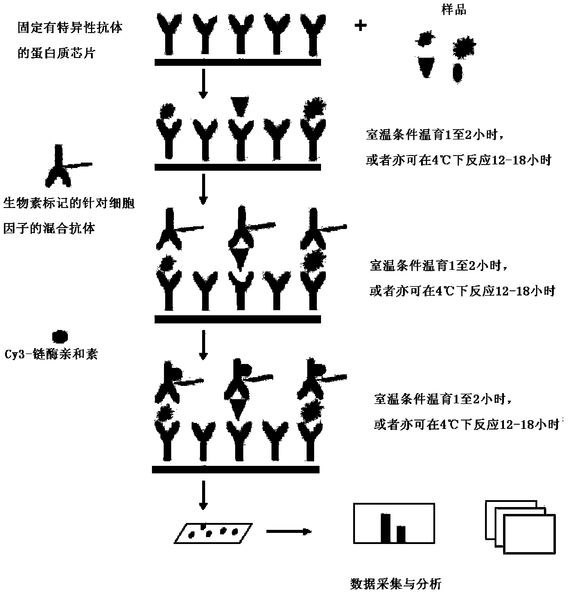
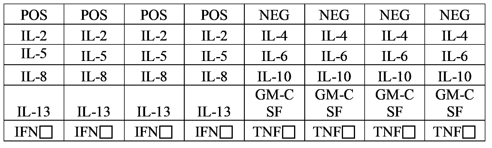
[0034] Example 2: Preparation of an antibody chip kit for simultaneous quantitative detection of multiple cytokines.
[0035] In order to detect the presence of the corresponding cytokines in the sample, prepare slides fixed with specific antibodies against the following proteins: granulocyte-macrophage colony stimulating factor (GM-CSF), interferon gamma (IFNgamma), interleukin-2 (IL-2), Interleukin 4 (IL-4), Interleukin 5 (IL-5), Interleukin 6 (IL-6), Interleukin 8 (IL-8), Interleukin 10 (IL-10), Interleukin 13 (IL -13), tumor necrosis factor alpha (TNFalpha).
[0036] 1. Preparation of antibody:
[0037] Using specific antibodies against the proteins listed in Table 1, the source, concentration, and names of the proteins against which the antibodies are directed are specified in Table 1:
[0038] Table 1 The name of the antigen protein targeted by the specific antibody, the source of the antibody, and the concentration information
[0039]
[0040] 2. Preparation and storage of antibody chip
[0041] Spot 100-1000pl of PBS buffer containing 0.02-2ng of specific antibody (containing 0.01-10g/100ml bovine albumin) on the glass slide with an automatic spotting instrument. The specific chip dot matrix diagram is as follows figure 2 As shown, biotin-labeled bovine IgG served as a positive control. There are also two positive controls of two different concentrations for each antibody, which are repeated four times in each chip. In this embodiment, the chip lattice uses such as figure 2 The arrangement shown, but in fact, in other embodiments, the chip dot matrix used for spotting can also be combined in other arrangements, and is not limited to the form shown in the figure. There are 16 identical chip dots on each slide. Place the spotted slides at room temperature overnight, and then dry them in a desiccator for 2 hours.
[0042] The dried slide is mounted on a specially designed 16-hole frame, and a slide is divided into 16 non-interfering cells. The frame structure is composed of three parts: the upper plastic hard frame, the lower soft latex film, and the plastic splints on both sides; the upper plastic hard frame and the lower soft latex film are fastened on the glass slide through the plastic splints on both sides to form The matrix is ​​a 2×8 16-well antibody chip; the size of each hole of the frame is consistent with the hole spacing of a conventional ELISA 96-well plate, so that it is suitable for the automated operation of a standard ELISA system. After the frame is sealed with an adhesive film, the entire chip is packaged in an airtight pouch and stored at 2°C to 8°C for later use.
[0043] In this embodiment, the automatic spotting instrument is a product produced by Bole Company or Platinum Elmer Company; the glass slide is a product of Corning Company of the United States. Of course, in the above steps of the technical solution of the invention, the use of instruments and materials is not limited to the enumeration of this embodiment, but is based on the ability to solve the technical problems of the invention and achieve corresponding technical effects.
[0044] 3. Preparation of cytokine standards:
[0045] The source, concentration, and name of the protein used for the recombinant proteins listed in Table 2 are detailed in Table 2:
[0046] Table 2: The name, source, and concentration information of the recombinant protein in the cytokine standard
[0047]
[0048] Dilute each of the above recombinant proteins with phosphate buffer containing 0.1% calf albumin and mix them together according to a certain amount. After packaging, they are dried by freeze-drying and stored at -80°C. In this example, the final concentration of each recombinant protein used for the standard curve is shown in Table 3. But in fact, in other embodiments, the concentration of recombinant protein used to make the standard curve can be selected in different intervals, which is not limited to the embodiment in Table 3.

Example Embodiment

[0049] Example 3: Experiment for quantitatively detecting cytokines using the kit of the present invention.
[0050] 1. Complete drying of the slide chip
[0051] Take the slide chip out of the box, after equilibrating at room temperature for 20-30 minutes, open the packaging bag, remove the sealing strip, and then place the chip in a vacuum desiccator or dry at room temperature for 1-2 hours.
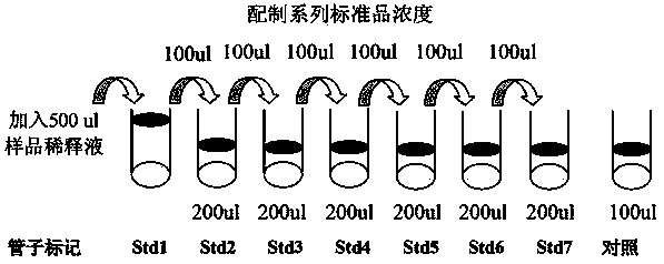
[0052] 2. Press image 3 Perform a serial dilution of the cytokine standard gradient as shown in
[0053] 2.1. Add 500μl of sample diluent to the small tube of cytokine standard mixture, and re-dissolve the standard. Before opening the vial, centrifuge it quickly, gently tap up and down to dissolve the powder, and mark this vial as Std1.
[0054] 2.2. Mark 6 clean centrifuge tubes as Std2, Std3 to Std7, and add 200μl of sample diluent to each tube.
[0055] 2.3. Extract 100μl of Std1 and add it to Std2 and mix gently, then extract 100μl from Std2 and add it to Std3, so as to gradually dilute to Std7.
[0056] 2.4. Take 100μl of sample dilution to another new centrifuge tube, labeled CNTRL, as a negative control.
[0057] Note: Because the initial concentration of each cytokine is different, the serial concentration of each cytokine is different after the gradient dilution of Std1 to Std7. In this example, the concentration of the gradient recombinant protein dilution is shown in Table 3. Shown.
[0058] Table 3: Concentrations of recombinant cytokine standards (pg/ml) used for standard curve after serial dilution
[0059]
[0060] 3. Chip operation process
[0061] 3.1. Add 100μl of sample diluent to each well, incubate for 30 minutes on a shaker at room temperature, and block the quantitative antibody chip.
[0062] 3.2. Remove the buffer from each well, add 100μl of standard solution and sample to the well, and incubate overnight at 4°C on a shaker.
[0063] Note: The amount of sample added for different samples is different: plasma and serum are diluted 1:1 with the sample diluent before use; the cell supernatant can be the original solution; the cell or tissue lysate is added to the amount of 50-500ug/ml after the protein concentration is determined .
[0064] 3.3, cleaning
[0065] Remove the standard or sample in each well, 1 wash 5 times from washing solution I, shake at room temperature for 5 minutes each time, 150μl of 1× washing solution I in each well, drain the washing solution every time and use it Dilute 20× lotion I with ionized water.
[0066] Remove 1× lotion I from each well, add 1× lotion II to wash twice, shake at room temperature for 5 min each time, 150μl 1× lotion II per well, clean the lotion every time. Dilute 20× lotion II with deionized water.
[0067] 3.4 Incubation of detection antibody mixture
[0068] Centrifuge the small tube of detection antibody mixture, then add 1.4ml of sample diluent, mix well and centrifuge again quickly. Add 80μl of detection antibody to each well and incubate for 2 hours on a shaker at room temperature.
[0069] 2.3.5 Cleaning
[0070] Remove the detection antibody from each well, wash 5 times with 1× Washing Solution I, shake at room temperature for 5 minutes each time, 150μl of 1× Washing Solution I in each well, draw clean wash solution for each wash, and then add 1× Wash the lotion II twice, shake at room temperature for 5 minutes each time, 150μl of 1× lotion II per well, drain the lotion every time.
[0071] 2.3.6 Incubation of Cy3-streptavidin
[0072] Centrifuge the Cy3-streptavidin vial, then add 1.4ml of sample diluent, mix well and centrifuge again quickly. Add 80μl of Cy3-streptavidin to each well, wrap the slide with aluminum foil and incubate in the dark, and incubate for 1 hour on a shaker at room temperature.
[0073] 3.7 Cleaning
[0074] Remove Cy3-streptavidin from each well, wash 5 times with 1× Washing Solution I, shaking at room temperature for 5 min each time, 150μl 1× Washing Solution I per well, and drain the washing solution every time. .
[0075] 3.8 Fluorescence detection
[0076] 1) Remove the frame of the slide, being careful not to touch the side of the slide with printed antibodies with your hands.
[0077] 2) Place the slide in the slide cleaning tube, add about 30ml of 1× Washing Solution I, which can cover the entire slide, shake for 15 minutes on a shaker at room temperature, discard 1× Washing Solution I, and add about 30ml of 1 × lotion II, shake for 5 min on a shaker at room temperature.
[0078] 3) Remove the residual lotion on the slide. Place the slides in the slide cleaning tube/drying tube without a lid, and centrifuge at 1000rpm for 3min.
[0079] 4) Use a laser scanner such as Axon GenePix to scan the signal, using Cy3 or green channel (excitation frequency = 532nm).
[0080] 2.3.9 Chip data extraction and analysis software for data analysis
[0081] 1) Use GenePix software to read the fluorescence value of the biochip. The microarray parameters of the chip are 6 (rows) x 8 (columns), and the dot diameter is 120um.
[0082] 2) The selected value after reading is the median reading (F532Median-Local Background) after removing the local background. Use the specific quantitative chip calculation software QAH-TH-1-SW to make the standard curve of each recombinant protein as shown in Figure 4.

PUM

no PUM

Description & Claims & Application Information

We can also present the details of the Description, Claims and Application information to help users get a comprehensive understanding of the technical details of the patent, such as background art, summary of invention, brief description of drawings, description of embodiments, and other original content. On the other hand, users can also determine the specific scope of protection of the technology through the list of claims; as well as understand the changes in the life cycle of the technology with the presentation of the patent timeline. Login to view more.

Similar technology patents

Classification and recommendation of technical efficacy words

  • Improve stability
  • Low background noise

Gel stabilized nanoparticulate active agent compositions

ActiveUS20050031691A1Improve stabilityImprove dissolutionPowder deliveryBiocideActive agentAverage diameter
Owner:ALKERMES PHARMA IRELAND LTD

Surgical ligature clip

InactiveUS20080312670A1Improve stabilityAvoid spaceWound clampsOrthodontic ligatureArm moving
Owner:AESCULAP AG

Electromechanical system equipment health assessment method

ActiveCN105930963AReduce test costLow background noiseResourcesHealth evaluationDecomposition
Owner:CSSC SYST ENG RES INST

Chemical phase analysis method for rubidium in metal ore

ActiveCN112179896ALow background noiseSimple methodAnalysis by thermal excitationAshingOxide
Owner:长沙矿冶院检测技术有限责任公司
Why Eureka
Try Eureka