Anti-tumor fusion protein, and preparation method and application thereof

A technology of fusion protein and prosthetic protein, applied in the field of biomedicine

Active Publication Date: 2014-10-29
MEDICINE & BIOENG INST OF CHINESE ACAD OF MEDICAL SCI +2
View PDF10 Cites 4 Cited by
  • Summary
  • Abstract
  • Description
  • Claims
  • Application Information

AI Technical Summary

Problems solved by technology

[0004] Although the combined use of vasopressor drugs such as endostar and chemotherapy drugs is a commonly used treatment in the field, there is no report on the combined use of endostatin and lidamycin in the field

Method used


Image

  • Anti-tumor fusion protein, and preparation method and application thereof
  • Anti-tumor fusion protein, and preparation method and application thereof
  • Anti-tumor fusion protein, and preparation method and application thereof

Examples


Embodiment 1

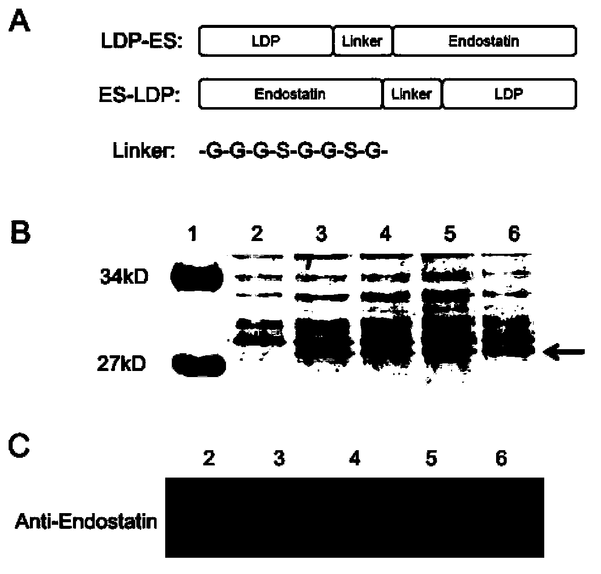
[0053]Embodiment 1, preparation recombinant expression construct

[0054] The DNA coding frame of the fusion protein composed of LDP and endostatin is prepared by PCR and molecular cloning techniques. The full-length coding sequence of fusion protein comprises the nucleotide sequence (SEQ ID NO: 8) of coding LDP and the nucleotide sequence (SEQ ID NO: 9) of coding endostatin, the sequence (SEQ ID NO: 9) by coding connecting peptide GGGSGGSG in the middle ID NO:10) separated. The obtained full-length coding sequence was inserted into the expression vector of pET30a to obtain an expression construct. like figure 1 As shown in A, two fusion proteins named ES-LDP (SEQ ID NO: 4) and LDP-ES (SEQ ID NO: 5) were constructed. The polynucleotide fragments (SEQ ID NO:6 and SEQ ID NO:7) encoding these two fusion proteins were respectively inserted into the pET30a expression vector, and then transformed into BL21(DE3) competent cells.

Embodiment 2

[0055] Example 2, expression, purification, renaturation and identification of fusion protein

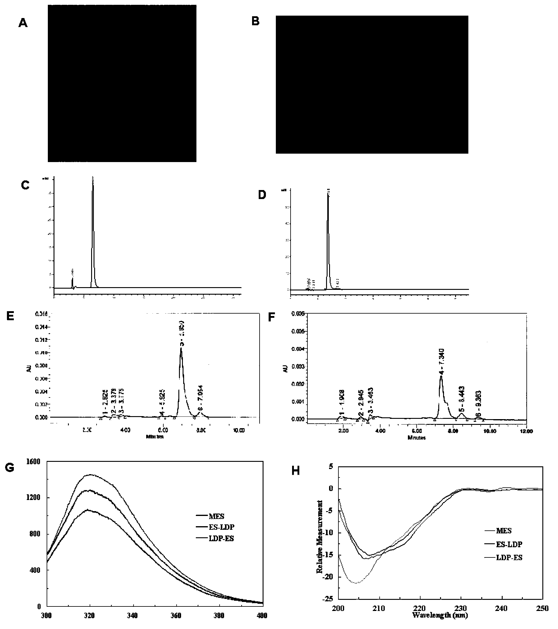
[0056] The transformed cells obtained in Example 1 were used to express the fusion protein on a large scale. by SDS-PAGE ( figure 1 B) and western blot ( figure 1 C) method to detect the expression of the fusion protein. The expressed fusion protein was purified and refolded for SDS-PAGE and HPLC analysis respectively. The result is as figure 2 Shown: figure 2 A represents the SDS-PAGE pattern of the fusion protein ES-LDP after renaturation after purification, in which: the left side is reducing electrophoresis; the right side is non-reducing electrophoresis; figure 2 B represents the SDS-PAGE profile of the fusion protein LDP-ES after renaturation after purification, in which: the left side is reducing electrophoresis; the right side is non-reducing electrophoresis; figure 2 C represents the RP-HPLC analysis of the fusion protein ES-LDP after renaturation, figure 2 D re...

Embodiment 3

[0057] Embodiment 3, the combination of fusion protein ES-LDP and LDP-ES and lidamycin chromophore

[0058] Get highly active lidamycin pure product (prepared and preserved by the Institute of Medical Biotechnology, Chinese Academy of Medical Sciences), and separate the active chromophore through a C4 column. The mobile phase is water: acetonitrile: trifluoroacetic acid (78%: 22% : 0.05%), monitor the absorbance at 350nm, and collect the lidamycin chromophore. The fusion protein obtained in Example 2 was mixed with the lidamycin chromophore at a molar ratio of 1:4, placed on a shaker and shaken slowly, and reacted at 4°C in the dark for 16 hours. The mixed solution was removed through the Sephadex G-25 column to remove the unbound lidamycin chromophore, and the absorbance value at 340nm was detected by HPLC (C4300A column) ( figure 2 E and F). The respective binding efficiencies of the two fusion proteins are calculated according to the standard curve of the lidamycin chrom...


PUM


Abstract

The invention discloses anti-tumor fusion protein, and a preparation method and application thereof. Concretely, the invention discloses fusion protein containing lidamycin apoprotein and endostatin, and the fusion protein can be reinforced by using lidamycin chromophore. The invention also discloses a method of generating the fusion protein through recombination and application of the fusion protein to treat tumor.

Description

technical field [0001] The invention belongs to the field of biomedicine, and relates to a fusion protein of endostatin and lidamycin prosthetic protein, a preparation method and application thereof. Background technique [0002] In 1971, Professor Folkman (Folkman J.N Engl J Med.1971;285:1182-1186) proposed that tumor growth depends on angiogenesis. New blood vessels in tumor tissue provide nutrients and oxygen for tumor growth, and are also the main pathway for tumor invasion and metastasis. Generally, benign tumors have sparse angiogenesis and slow-growing blood vessels; whereas most malignant tumors have dense angiogenesis and grow rapidly. Therefore, inhibition of angiogenesis can significantly prevent tumor growth and spread. In recent years, new anticancer drugs targeting tumor angiogenesis (such as bevacizumab, sorafenib, sunitinib) have entered clinical application and achieved good curative effect. In addition, as a broad-spectrum inhibitor of endogenous angioge...

Claims


Application Information

Patent Timeline
Patent Type & Authority Applications(China)
IPC IPC(8): C07K19/00C12N15/62C12N15/63C12N1/21C12N1/19C12N1/15C12P21/00A61K38/16A61P35/00C12R1/19C12R1/465C12R1/38
Inventor 甄永苏罗永章姜文国鲁薪安尚伯杨付彦张胜华周代福李良李毅
Owner MEDICINE & BIOENG INST OF CHINESE ACAD OF MEDICAL SCI
Why Eureka
Try Eureka