Method for preparing intravascular stent upon 3D (three-dimensional) printing technology

A 3D printing, 3D printer technology, applied in brackets and other directions, can solve problems such as hazards, microstructure defects, stepped surfaces, etc., to achieve the effects of cost reduction, good biocompatibility, and complete surface

Active Publication Date: 2014-12-24
HUNAN HANDLIKE MINIMALLY INVASIVE SURGERY CO LTD
View PDF7 Cites 20 Cited by
  • Summary
  • Abstract
  • Description
  • Claims
  • Application Information

AI Technical Summary

Problems solved by technology

However, the preparation of medical devices generally has relatively high requirements on materials. The materials produced by 3D printing are often due to the selection of different materials, inappropriate binders selected during molding, and poor control of process parameters, which will lead to blanks. Defects such as fuzzy body surface, warping deformation, size deformation, stepped surface, microstructure defects, fragmentation, and mislayering are fatal hazards for the use of 3D printing technology in the manufacture of medical devices
Moreover, due to the shrinkage and deformation of the product during the sintering process, it is difficult to control the dimensional accuracy of the product.

Method used


Image

  • Method for preparing intravascular stent upon 3D (three-dimensional) printing technology
  • Method for preparing intravascular stent upon 3D (three-dimensional) printing technology

Examples


Embodiment 1

[0045] Step (1): Modeling

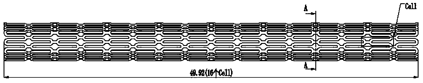
[0046] Establishing a 3D model of the intravascular stent in a computer, and decomposing the 3D model into a series of two-dimensional thin slice models with a thickness of 90 μm;

[0047] Step (2): Ingredients

[0048] Powder material: The stainless steel powder material is made by mixing spherical 316L stainless steel powder with an average particle size of 8 μm and stearic acid powder at a volume ratio of 60:40 at a temperature of 140°C;

[0049] Binder: a paraffin-based binder is made by mixing paraffin, low-density polyethylene, polypropylene and stearic acid in a mass ratio of 68:23:8:1;

[0050] Step (3): 3D printing to prepare green body

[0051] Input the model data built in step (1) into the supporting equipment of the 3D printer to set the printing program, send the stainless steel powder material in step (2) to the printing platform of the 3D printer, roll the layer, and the print head of the 3D printer The paraffin-based binder in th...

Embodiment 2

[0056] Step (1): Modeling

[0057] Establishing a 3D model of the intravascular stent in a computer, and decomposing the 3D model into a series of two-dimensional thin slice models with a thickness of 100 μm;

[0058] Step (2): Ingredients

[0059] Powder material: The stainless steel powder material is made by mixing spherical 316L stainless steel powder with an average particle size of 15 μm and stearic acid powder at a volume ratio of 80:20 at a temperature of 155°C;

[0060] Binder: a paraffin-based binder is made by mixing paraffin wax, low-density polyethylene, polypropylene and stearic acid in a mass ratio of 70:20:9:1;

[0061] Step (3): 3D printing to prepare green body

[0062] Input the model data built in step (1) into the supporting equipment of the 3D printer to set the printing program, send the stainless steel powder material in step (2) to the printing platform of the 3D printer, roll the layer, and the print head of the 3D printer The paraffin-based binder...

Embodiment 3

[0067] Step (1): Modeling

[0068] Establishing a 3D model of the intravascular stent in a computer, and decomposing the 3D model into a series of two-dimensional thin slice models with a thickness of 85 μm;

[0069] Step (2): Ingredients

[0070] Powder material: The nickel-titanium powder material is made by mixing spherical nickel-titanium powder with an average particle size of 10 μm and stearic acid powder at a volume ratio of 70:30 at a temperature of 145°C;

[0071] Adhesive: a cyano-adhesive with a mass percentage concentration of 0.8% made from α-ethyl cyanoacrylate dissolved in water;

[0072] Step (3): 3D printing to prepare green body

[0073] Input the model data built in step (1) into the supporting equipment of the 3D printer to set the printing program, send the nickel-titanium powder material in step (2) to the printing platform of the 3D printer, roll the layer, and print the 3D printer The cyano-based binder in the head spraying step (2) sticks the nickel...


PUM

PropertyMeasurementUnit
thicknessaaaaaaaaaa
particle sizeaaaaaaaaaa

Abstract

The invention discloses a method for preparing an intravascular stent upon the 3D (three-dimensional) printing technology. The method comprises the following steps: firstly establishing a 3D model of the intravascular stent in a computer; inputting data of the established model into corollary equipment of a 3D printer and setting a printing program; bonding a mixed powder material, consisting of stainless steel powder or nickel-titanium powder and stearic acid powder, with a bonder through 3D printing program control to form an intravascular stent blank; performing degreasing, vacuum sintering and cooling treatment on the blank in sequence to obtain the intravascular stent. According to the preparation method, the personalized model can be designed according to the actual requirement of a patient, and the required intravascular stent blank can be rapidly and accurately prepared; the further prepared intravascular stent has a complete surface and has no defects of deformation or cracking and the like; especially, the prepared intravascular stent has no rejection reaction in 24 days in simulated body fluid, is good in biocompatibility and completely meets the medical requirement; the cost for preparing the intravascular stent by a traditional laser cutting method is greatly reduced.

Description

technical field [0001] The invention relates to a method for preparing an intravascular stent based on 3D printing technology, which belongs to the field of medical device preparation. Background technique [0002] Vascular stent technology is a minimally invasive interventional treatment method, which is used to support narrowed and occluded blood vessels in the human body due to disease, and to restore blood circulation. primary means. The processing material of the vascular stent should be corrosion-resistant, have good compatibility with the vessel wall and blood, be fatigue-resistant, and can be clearly visualized under X-rays. At present, the widely used intravascular stent is mainly prepared by tube laser cutting process, but this method is costly, the design of the stent structure is limited, and it is difficult to realize the complex shape of the vascular stent, and the key performance such as accuracy and smoothness cannot be achieved, so it is in clinical practic...

Claims


Application Information

Patent Timeline
Patent Type & Authority Applications(China)
IPC IPC(8): A61F2/90
Inventor 舒畅李益民何浩王暾赵志刚张翔陈至杨
Owner HUNAN HANDLIKE MINIMALLY INVASIVE SURGERY CO LTD
Why Eureka
Try Eureka