Method for detecting external capsule bodies in liquid sample

A technology of liquid samples and exocysts, which is applied in the direction of measuring devices, scattering characteristics measurement, instruments, etc., can solve the problems of negative impact on the credibility of test results, labeling technology increases the background noise of non-specific interaction detection, etc.

Active Publication Date: 2014-12-24
THE NAT CENT FOR NANOSCI & TECH NCNST OF CHINA
View PDF7 Cites 0 Cited by
  • Summary
  • Abstract
  • Description
  • Claims
  • Application Information

AI Technical Summary

Problems solved by technology

Although there is a recent method based on immunomagnetic beads to directly detect exocysts in cell culture supernatants, this method still needs to be labeled with secondary antibodies such as HRP for functional studies, and the labeling technique is prone to increase non-specificity Interaction and detection background noise, which negatively affects the confidence of detection results

Method used


Image

  • Method for detecting external capsule bodies in liquid sample
  • Method for detecting external capsule bodies in liquid sample
  • Method for detecting external capsule bodies in liquid sample

Examples


preparation example Construction

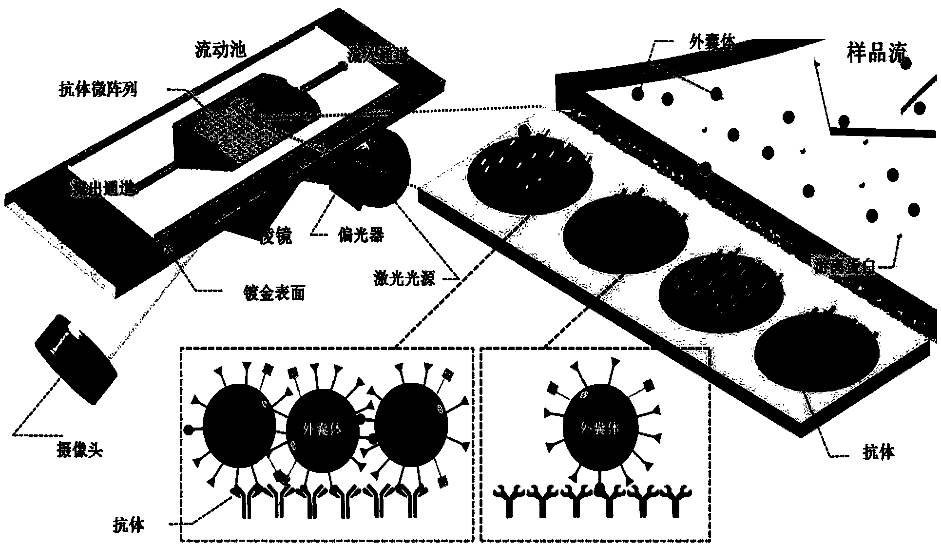
[0020] The preparation method of the microarray chip loaded with exosome surface membrane protein specific antibody according to the present invention can be according to the literature (Lausted, C.; Hu, Z.; Hood, L.; Campbell, C.T. Comb Chem High Throughput Screen 2009 , 12, (8), 741-51.), preferably, the preparation process may include the following steps:

[0021] Printing: Print one or more specific antibodies to exocyst surface membrane proteins at a concentration of 0.1-10 μg / μL on a chip coated with a metal layer on the surface to form an antibody microarray. The chip can be a gold-plated chip or a silver-plated chip commonly used in SPRi technology. The size of the chip can be the size conventionally used in the art. Generally, the printing density of antibodies on the chip per square centimeter is 1-10000 dots, preferably 1-100 dots. In terms of protein amount, each The amount of antibody in the antibody spot is 0.02-2 μg, preferably 0.1-1 μg. After printing, place ...

preparation example 1

[0056] This preparation example is used to illustrate the preparation method of the microarray chip provided by the present invention. In this preparation example, the same microarray chips 1-6 are prepared for subsequent test examples. The specific preparation steps are:

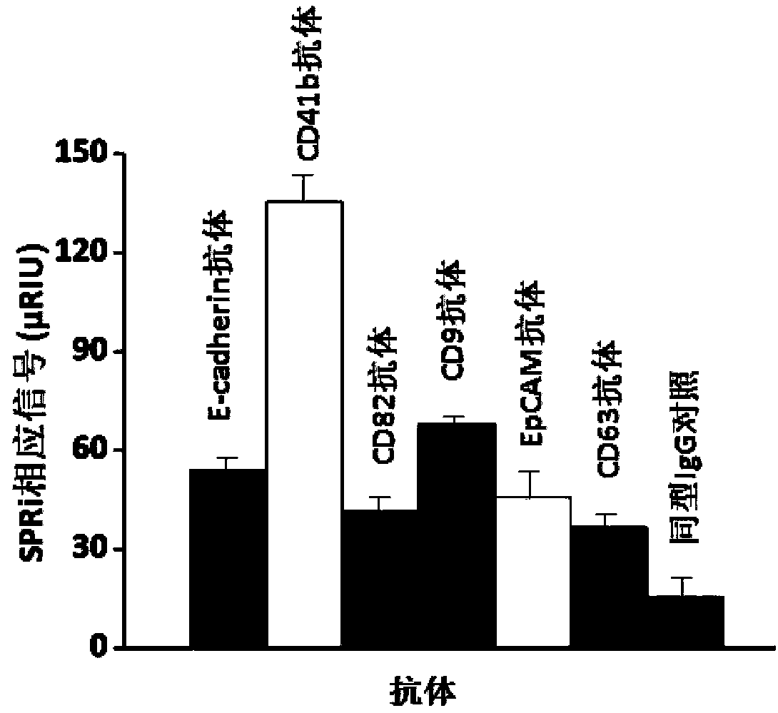
[0057] (1) Printing: Mouse anti-CD63 IgG, mouse anti-CD9 IgG, mouse anti-CD41b IgG, mouse anti-E-cadherin IgG, mouse anti-EpCAM IgG, mouse anti-CD82 IgG and the control group IgG at a concentration of 5 μg / μL at 5000 cells / cm 2 The printing density was fixed to the surface of the plasmon resonance imaging gold-plated chip modified by polyethylene glycol to form an antibody microarray, and one spot was printed for each antibody (the amount of protein in each spot was 0.5 μg), and the microarray chip was placed Incubate at 40°C for 2 hours in a humid box with a humidity of 50%;

[0058] (2) Cleaning: Wash the chip with 100ml of 1M high-fold phosphate buffer, 100ml of 0.1M low-fold phosphate buffer and 200ml ...


PUM

PropertyMeasurementUnit
diameteraaaaaaaaaa
diameteraaaaaaaaaa

Abstract

The invention provides a method for detecting external capsule bodies in a liquid sample. The liquid sample to be detected contains the external capsule bodies. The method comprises the steps of feeding the liquid sample to be detected into the surface of a microarray chip for being in contact with each other, wherein the surface of the microaray chip is loaded with one or more kinds of specific antibodies with external capsule body surface membrane proteins so as to obtain surface plasma resonance signal change values before and after the microarray chip is in the contact with the liquid sample to be detected; and indicating the quantity of the external capsule bodies in the liquid sample to be detected according to the surface plasma resonance signal change values of the microarray chip. The method provided by the invention is based on a surface plasma resonance imaging technology, so that the liquid sample to be detected does not need to be subjected to complicated purification, and the external capsule bodies in the liquid sample to be detected can be accurately, reliably and quantitatively detected in real time.

Description

technical field [0001] The invention relates to a method for quantitatively detecting exocysts in liquid samples. Background technique [0002] Surface plasmon resonance (SPR) sensing technology is a surface-sensitive detection method. Compared with other biological detection methods, the SPR sensing analysis system has the advantages of label-free and real-time detection of interaction kinetics. . The surface plasmon resonance imaging (SPRi) analysis system based on SPR technology not only maintains the advantages of SPR detection technology, but also increases the detection capacity. SPRi can detect hundreds or even thousands of interactions at the same time. A high-throughput detection technique. [0003] The exosome is a flat spherical microvesicle surrounded by a phospholipid bilayer with a diameter in the range of 40-100 nm. The formation process of exocysts is relatively complicated. At present, scientific researchers generally believe that exocysts originate from ...

Claims


Application Information

Patent Timeline
Patent Type & Authority Applications(China)
IPC IPC(8): G01N21/55G01N33/564
Inventor 胡志远崔健王坤
Owner THE NAT CENT FOR NANOSCI & TECH NCNST OF CHINA
Why Eureka
Try Eureka