Surface enhanced Raman spectra substrate for detecting avian influenza virus, and application thereof

A surface-enhanced Raman and avian influenza virus technology, applied in Raman scattering, material excitation analysis, etc., can solve the problems of low virus content, complex sample components, false negatives, etc., and achieve the effect of rapid detection and identification

Active Publication Date: 2015-03-11
INST OF MICROBIOLOGY - CHINESE ACAD OF SCI +1
View PDF4 Cites 10 Cited by
  • Summary
  • Abstract
  • Description
  • Claims
  • Application Information

AI Technical Summary

Problems solved by technology

Surface-enhanced Raman spectroscopy can be applied to the detection of avian influenza virus. However, since the clinical samples to be detected are mostly feces or cloacal swabs, the sample composition is complex, and the virus content is low, so the miscellaneous peaks of the SERS spectrum cover the effective peaks. , resulting in a false negative result

Method used


Image

  • Surface enhanced Raman spectra substrate for detecting avian influenza virus, and application thereof
  • Surface enhanced Raman spectra substrate for detecting avian influenza virus, and application thereof
  • Surface enhanced Raman spectra substrate for detecting avian influenza virus, and application thereof

Examples


Embodiment 1

[0048] Example 1. Preparation of surface-enhanced Raman spectroscopy substrate for detection of avian influenza virus

[0049] 1. Preparation of gold nanoparticles

[0050] 50mg of chloroauric acid is dissolved in 500ml of ultrapure water, after heating and boiling, add 1% (1g / 100ml) trisodium citrate solution (solvent is water) of 8.7ml, stir vigorously and continue to boil and cool to room temperature after 35min ( 25° C.) to obtain gold nanoparticles with a diameter of about 10-15 nm.

[0051] 2. Preparation of gold nanoparticles coupled with α(2,3) sialic acid oligosaccharides

[0052] 1. Preparation of α(2,3) sialotriose-short connecting chain

[0053] For the specific preparation method, refer to Example 1 of the Chinese patent application with application number 201310495249.x (publication number CN 103551562A). The α(2,3) sialotriose-short connecting chain as shown in formula III below and the short connecting chain as shown in formula III are obtained.

[0054] Fo...

Embodiment 2

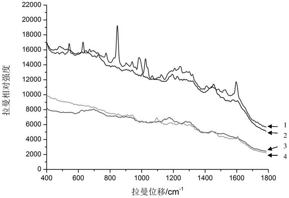
[0059] Example 2, Verification of Surface Enhanced Raman Spectroscopy Base Substrate Detection of Avian Influenza Virus

[0060] The gold nanoparticles coupled with α(2,3) sialic acid oligosaccharides obtained in Example 1 were used as a substrate, mixed with different influenza virus samples, and detected by surface-enhanced Raman spectroscopy, and simultaneously set up without adding virus samples. The detection control containing only the substrate, and the non-conjugated sialooligosaccharide gold nanoparticles in Example 1 were selected for the detection control. The specific implementation plan is as follows:

[0061] 1. Take four 1.5ml centrifuge tubes, and add 100 μl of the nano-gold solution coupled with α(2,3) sialic acid oligosaccharides obtained in Example 1 to the three centrifuge tubes, numbered 1, 3, 4, and the other Add 100 μl of unconjugated sialooligosaccharide gold nanoparticles, numbered 2, to the centrifuge tube. Then add 10 μl avian influenza virus A / Vie...

Embodiment 3

[0064] Example 3, Application of Surface Enhanced Raman Spectroscopy Substrates to Detect Different Subtypes of Avian Influenza Viruses

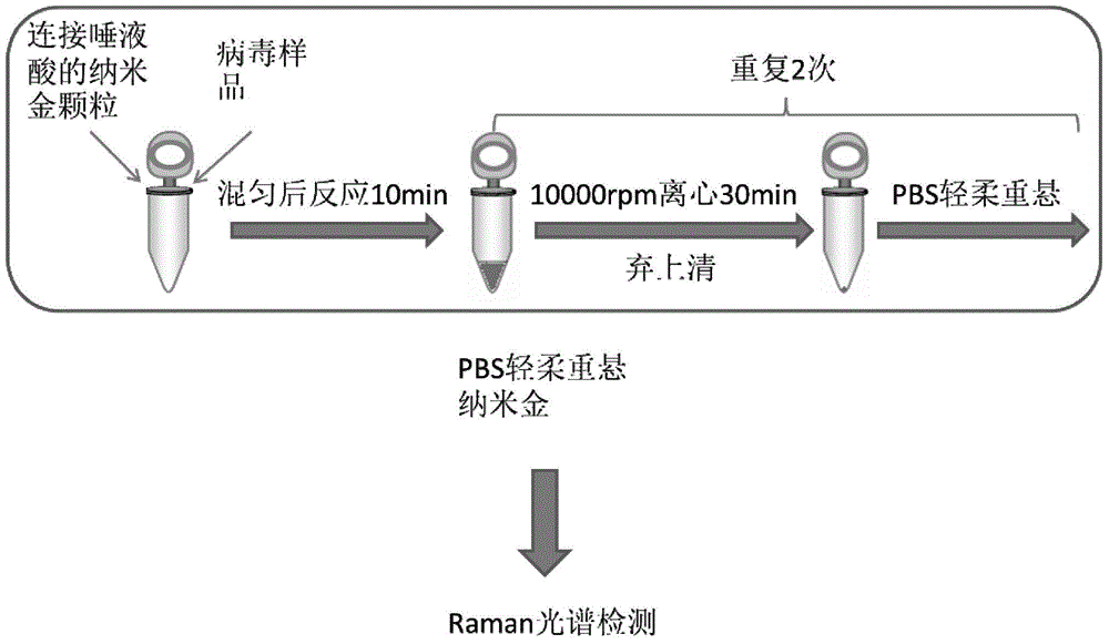
[0065] The gold nanoparticles coupled with α(2,3) sialic acid oligosaccharides obtained in Example 1 are mixed with different influenza virus strain samples respectively as substrates, and then surface-enhanced Raman spectroscopy detection is carried out. The operation flow chart is as follows figure 2 shown. The specific implementation plan is as follows:

[0066] 1. Take four 1.5ml centrifuge tubes, add 100μl of nano-gold solution coupled with α(2,3) sialic acid oligosaccharides, numbered 1, 2, 3, 4, and then add 10μl poultry Influenza virus A / Chicken / Shanghai / 441 / 2009 (H9N2) strain samples (10 7 Virus particles / ml), No. 2 tubes add 10 μl avian influenza virus A / Vietnam / 1194 / 2004 (H5N1) strain sample (10 7 Virus particles / ml), No. 3 tubes add 10 μ l avian influenza virus A / Puerto Rico / 8 / 34 (H1N1) strain samples (10 7 Virus particles / m...


PUM

PropertyMeasurementUnit
diameteraaaaaaaaaa
diameteraaaaaaaaaa
particle diameteraaaaaaaaaa

Abstract

The invention discloses a surface enhanced Raman spectra substrate for detecting an avian influenza virus and application of the surface enhanced Raman spectra substrate. The application provided by the invention specifically relates to the application of nano-gold particles coupled with alpha (2 and 3) sialyloligosaccharide in the surface enhanced Raman spectra substrate for detecting the avian influenza virus. Experiments show that the substrate can specifically capture the avian influenza virus in a sample to be detected; the enrichment of avian influenza viruses in the sample can be realized through centrifugal collection, and then the enriched avian influenza viruses are used for SERS (Surface Enhanced Raman Scattering) spectra detection. An interference wave crest caused by sample impurities is effectively removed because of the enrichment of the viruses, and a spectral signal can also be effectively amplified by the nano-gold particles, so that the substrate can be used for the SERS spectra detection of the avian influenza virus. The substrate has a great significance to the rapid detection and identification of the avian influenza virus.

Description

technical field [0001] The invention belongs to the field of virus detection, and relates to a surface-enhanced Raman spectrum substrate for detecting bird flu virus and an application thereof. Background technique [0002] In 1928, Indian physicist C.V.Raman discovered that when monochromatic light is incident on a sample, the molecules in the sample will scatter the incident light. In addition to the spectral lines with the same frequency as the incident light, the scattered light also has shifted spectral lines. , we call this kind of scattering Raman (Raman) scattering. Raman spectroscopy belongs to the category of molecular rotation and vibration spectroscopy, and most of the existing molecular structure information is obtained from molecular spectroscopy. Therefore, Raman spectroscopy has important application value in physics, chemistry, life science, medicine, material science and other fields. However, the spontaneous Raman scattering signal is very weak, thus lim...

Claims


Application Information

Patent Timeline
Patent Type & Authority Applications(China)
IPC IPC(8): G01N21/65
Inventor 刘文军李学兵杨利敏李晶贾晓娟孙蕾
Owner INST OF MICROBIOLOGY - CHINESE ACAD OF SCI
Why Eureka
Try Eureka