Injectable PLGA porous composite microsphere preparation embedded with BMP-2 containing particles and preparation method and application thereof

A technology of porous composite microspheres and BMP-2, which is applied in the field of medicine to avoid burst release and improve curative effect

Inactive Publication Date: 2015-03-25
SECOND MILITARY MEDICAL UNIV OF THE PEOPLES LIBERATION ARMY
View PDF5 Cites 14 Cited by
  • Summary
  • Abstract
  • Description
  • Claims
  • Application Information

AI Technical Summary

Problems solved by technology

[0006] At present, there is no literature reporting a porous microsphere preparation that has a 3D porous structure that is conducive to the attachment and growth of seed cells, and can release BMP-2 in a sustained and controlled manner, and can be used for injection.

Method used


Image

  • Injectable PLGA porous composite microsphere preparation embedded with BMP-2 containing particles and preparation method and application thereof
  • Injectable PLGA porous composite microsphere preparation embedded with BMP-2 containing particles and preparation method and application thereof
  • Injectable PLGA porous composite microsphere preparation embedded with BMP-2 containing particles and preparation method and application thereof

Examples


Embodiment 1

[0072] Example 1: Preparation of PLGA porous composite microspheres containing bone morphogenetic protein-2 (BMP-2) chitosan microspheres

[0073] A, prepare chitosan microspheres containing BMP-2:

[0074] According to literature reports (Kim SE, Park JH, Cho YW, Chung H, Jeong SY, Lee EB, et al. Porous chitosan scaffold containing microspheres loaded with transforming growth factor-beta1: implications for cartilage tissue engineering. Journal of controlled release: official journal of the Controlled Release Society.2003;91:365-74), prepared by the following method: weigh 120mg chitosan (molecular weight 100000, Zhejiang Jinke Biochemical Co., Ltd.), add 3mL 2% (V / V) acetic acid aqueous solution After vortexing to dissolve, add 1mL of 1% (W / V) BMP-2 (Peprotech, USA) aqueous solution, mix well and drop into 90mL n-octanol (containing 4% Span 80, V / V) After stirring at 1200 rpm for 30 minutes, 10 mL of 5% (W / V) sodium tripolyphosphate (Sodium tripopolyphosphate, TPP, American ...

Embodiment 2

[0077] Embodiment 2: Morphological observation of PLGA porous composite microspheres embedded with BMP-2 chitosan microspheres

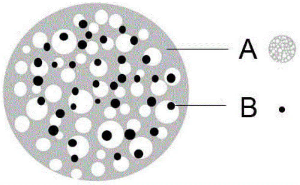
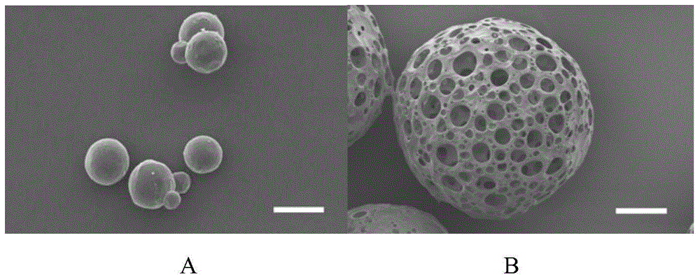
[0078] Get a certain amount of BMP-2 chitosan microspheres prepared in Example 1 and the PLGA porous composite microspheres containing BMP-2 chitosan microspheres, place on the sample plate that is glued with double-sided adhesive, gold-plated After observation under scanning electron microscope (ZESS MA10, Germany), chitosan microspheres ( figure 2 A, the bar is 20 μm) and PLGA porous composite microspheres containing BMP-2 chitosan microspheres ( figure 2 B, Scale bar is 100 μm) Morphology.

[0079] Chitosan microspheres are round in shape and smooth in surface, such as figure 2 As shown in A. The shape of PLGA porous microspheres is round, and a large number of pores are randomly distributed on the surface, such as figure 2 Shown in B.

Embodiment 3

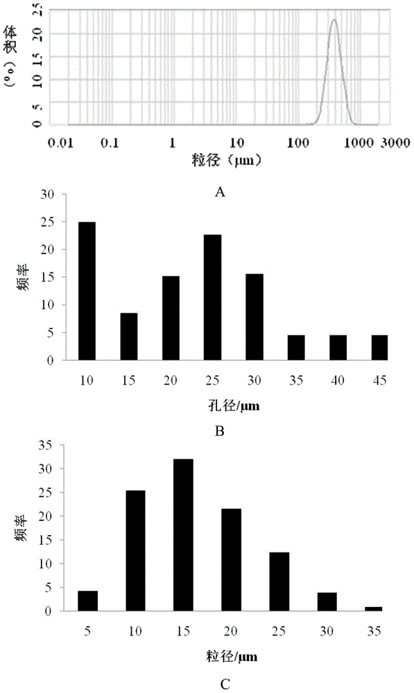
[0080] Embodiment 3: the particle size determination of the PLGA porous composite microspheres embedded with BMP-2 chitosan microspheres

[0081] The PLGA porous composite microspheres containing the BMP-2 chitosan microspheres inlaid prepared in Example 1 are dispersed in deionized water, and their particle diameter is measured with Malvern Masersizer2000 ( image 3 A). Randomly select 3 microspheres from the scanning electron microscope photos of porous microspheres, measure their surface pore size (negligible if the pore size is less than 5 μm), and take the average value. The pore size distribution diagram is as follows: image 3 Shown in B. Measure the particle diameter of the microspheres on the scanning electron microscope photos containing BMP-2 chitosan microspheres, and get the average value ( image 3 C).

[0082] Such as image 3 As shown in A, the average particle size of the PLGA porous composite microspheres embedded with BMP-2 chitosan microspheres is 454.0...


PUM

PropertyMeasurementUnit
particle sizeaaaaaaaaaa
pore sizeaaaaaaaaaa
particle sizeaaaaaaaaaa

Abstract

The invention belongs to the technical field of medicine, and provides an injectable PLGA porous composite microsphere preparation embedded with BMP-2 containing particles. A drug-carrying particle is formed by wrapping BMP-2 with hydrophilic materials such as chitosan, collagen or albumin; a microsphere with a 3D porous structure is constructed by the drug-carrying particle and a porous microsphere material PLGA; the drug-carrying particle is embedded inside the porous microsphere. The invention also provides a preparation method of the porous composite microsphere preparation, and an application in preparation of a repair material for defects of bone or cartilage tissue. The porous composite microsphere preparation of the invention can not only realize slow release of drugs at an administration site, but also facilitate proliferation and differentiation of porous supports of seed cells.

Description

technical field [0001] The invention belongs to the technical field of medicine, and in particular relates to an injectable PLGA porous composite microsphere preparation inlaid with BMP-2 microparticles and its preparation method and application. Background technique [0002] Bone and cartilage damage caused by trauma and pathology is a huge challenge for orthopedic surgeons. Conventional surgical treatment often brings unbearable pain to patients, and the treatment effect is unpredictable, often leading to nonunion . Using bone tissue engineering, the use of degradable polymer materials to make tissue engineering scaffolds to repair bone and cartilage damage is a research hotspot in recent years. Tissue engineering is a discipline that applies the principles and technologies of life science and engineering to study biological substitutes for repairing, maintaining and promoting the function and shape of human tissues or organs after damage. With the need of non-invasive a...

Claims


Application Information

Patent Timeline
Patent Type & Authority Applications(China)
IPC IPC(8): A61L27/20A61L27/22A61L27/18A61L27/50A61L27/56A61L27/54C08J9/26C08J9/28
Inventor 陈琰钟延强陶春黄景斌邹豪鲁莹刘俊杰张翮俞媛高静孙治国
Owner SECOND MILITARY MEDICAL UNIV OF THE PEOPLES LIBERATION ARMY
Why Eureka
Try Eureka