

Magnetic resonance imaging method of magnetization transfer and level choose inversion recovery combined pre-pulse
A magnetic resonance imaging and inversion recovery technology, applied in medical science, sensors, diagnostic recording/measurement, etc., can solve the problems of inability to obtain satisfactory suppression effect, prolonged signal acquisition time, poor background suppression effect, etc., to make up for inspection Long time, easy to use, improve the effect of vascular inflow enhancement effect
- Summary
- Abstract
- Description
- Claims
- Application Information
AI Technical Summary
Problems solved by technology
Method used
Image

Examples
Embodiment Construction
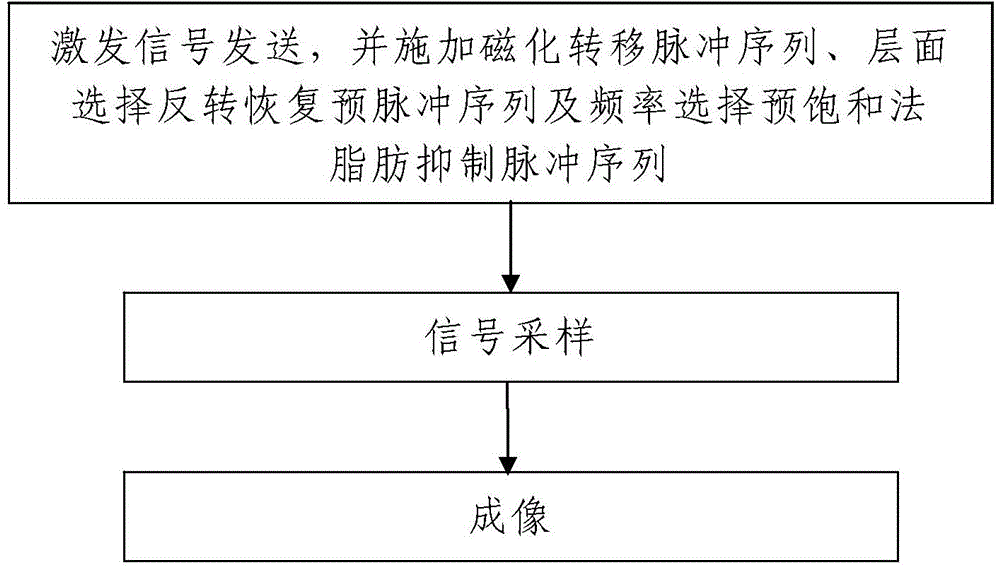
[0043] Such as figure 1 A magnetic resonance imaging method of magnetization transfer combined with layer-selective inversion recovery pre-pulse is shown, which uses a magnetic resonance imaging device to perform non-contrast agent-enhanced angiography under free breathing, and the method includes the following steps:
[0044] Step 1. Sending excitation signals: using the magnetic resonance imaging equipment to send excitation signals to the detected object, and the transmitted excitation signals are scanning imaging sequences.
[0045] When sending the excitation signal, the magnetic resonance imaging equipment is used to apply the magnetization transfer pulse sequence and the slice selective inversion recovery pre-pulse sequence according to the combination of magnetization transfer and layer selective inversion recovery.
[0046] Step 2. Signal sampling: the magnetic resonance imaging equipment is used to sample the magnetic resonance data of the detected object, and the ma...
PUM
 Abstract
Description
Claims
Application Information