17beta-estradiol visualization detection method based on DNA nano-structure, and 17beta-estradiol visualization detection kit based on DNA nano-structure

A detection kit and nanostructure technology, applied in the field of analytical chemistry, can solve the problems of high detection cost, troublesome storage, limited wide application, etc., and achieve the effects of high sensitivity, simple operation and good specificity

Active Publication Date: 2015-10-14
GUANGDONG INST OF ECO ENVIRONMENT & SOIL SCI
View PDF0 Cites 20 Cited by
  • Summary
  • Abstract
  • Description
  • Claims
  • Application Information

AI Technical Summary

Problems solved by technology

Traditional 17β-estradiol detection methods mainly include high performance liquid chromatography, gas chromatography-mass spectrometry, liquid chromatography-mass spectrometry, etc. These methods require cumbersome sample pretreatment and expensive instruments, and the detection cost is high, time-consuming and labor-intensive, and requir...

Method used


Image

  • 17beta-estradiol visualization detection method based on DNA nano-structure, and 17beta-estradiol visualization detection kit based on DNA nano-structure
  • 17beta-estradiol visualization detection method based on DNA nano-structure, and 17beta-estradiol visualization detection kit based on DNA nano-structure
  • 17beta-estradiol visualization detection method based on DNA nano-structure, and 17beta-estradiol visualization detection kit based on DNA nano-structure

Examples


Embodiment 1

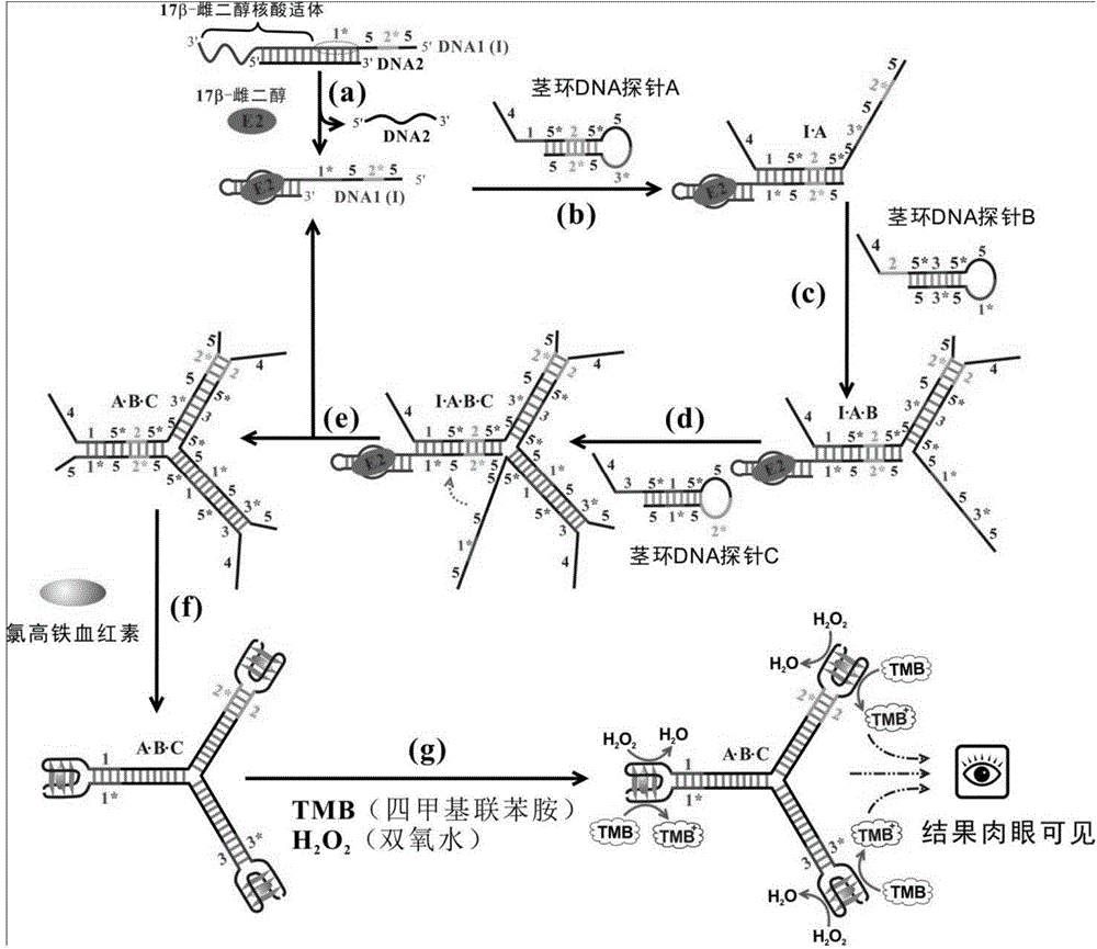
[0050] A 17β-estradiol visualization detection kit based on DNA nanostructure, which comprises the following components:

[0051] (1) DNA1:

[0052] 52*51*17β-estradiol nucleic acid aptamer

[0053] 5'-ATGGGT—CTCACT--ATGGGT--TCAACG—GCTTCCAGCTTATTGAATTACACGCAGAGGGTAGCGGCTCTGCGCATTCAATTGCTGCGCGCTGAAGCGCGGAAGC-3' (SEQ ID NO. 1)

[0054] The 5' or 3' end of the 17β-estradiol aptamer was extended to form DNA1, which included 5 units, 2* units, 5 units, 1* units and the 17β-estradiol aptamer region.

[0055] (2) DNA2:

[0056] 5'-ATTCAATAAGCTGGAAGCCGTTGA-3' (SEQ ID NO.2)

[0057]DNA2 is complementary to the 1* region of DNA1 and part of the 17β-estradiol aptamer region.

[0058] (3) Stem-loop DNA probe A:

[0059] 415*25*53*

[0060] 5'-TGGGTAGGGCGGGT--CGTTGA-ACCCAT--AGTGAG-ACCCAT-ATGGGT--CAAGAC--ATGGGT--CTCACT--ATGGGT-3' (SEQ ID NO. 3)

[0061] 52*5

[0062] Stem-loop DNA probe A comprises 4 units, 1 unit, 5* unit, 2 units, 5* units, 5 units, 3* units, 5 units, 2* units and...

Embodiment 2

[0089] Utilize the kit established in Example 1 to detect 17β-estradiol, the steps are as follows:

[0090] (1) First use Tris-HCl buffer (20mM, pH 7.4, containing 200mMNaCl and 50mMKCl) to dissolve DNA1, DNA2 and stem-loop DNA probes respectively;

[0091] (2) Mix 100 nM DNA1 and 400 nM DNA2, and react at room temperature for 20 minutes to form a DNA1-DNA2 mixture;

[0092] (3) Add the sample to be tested into the DNA1-DNA2 mixture, and react at room temperature for 45 minutes;

[0093] (4) Then add 1 μM stem-loop DNA probes A, B, and C, and react at room temperature for 60 minutes;

[0094] (5) Add 0.3 μM hemin (hemin) and react at room temperature for 30 minutes;

[0095] (6) Take 50 μL of the reaction solution and add it to 950 μL of chromogenic buffer (containing 26.6 mM citric acid, 51.4 mM disodium hydrogen phosphate, 25 mM KCl, 10 μL of 0.5% TMB, 20 μL of 30% H 2 o 2 , pH=5.0), reacted at room temperature for 15 minutes, and observed the color change;

[0096] If ...

Embodiment 3

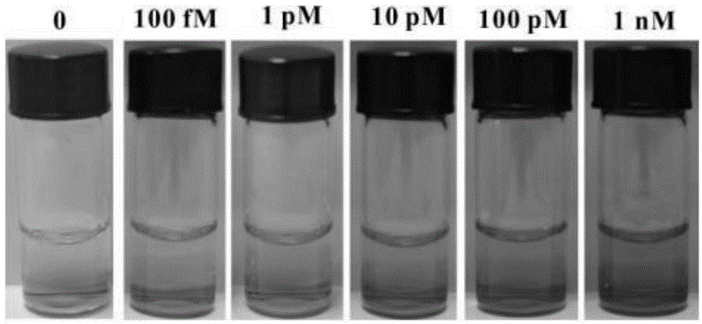
[0098] Detection of different concentrations of 17β-estradiol:

[0099] Prepare 17β-estradiol standard solutions with concentrations of 100fM, 1pM, 10pM, 100pM and 1nM respectively, and store at room temperature.

[0100] 17β-estradiol solutions of different concentrations were added to the reaction system described in Example 1 respectively, and the experimental results were observed after sufficient reaction, such as figure 2 As shown, 100fM of 17β-estradiol can produce obvious blue color change, indicating that its detection limit is 100fM. As the concentration of 17β-estradiol increased, the color also increased and gradually became saturated.


PUM


Abstract

The present invention discloses a 17beta-estradiol visualization detection method based on DNA nano-structure, and a 17beta-estradiol visualization detection kit based on DNA nano-structure. The working principle is that 17b-estradiol interacts with aptamer, a strand displacement reaction is started, three groups of stem-loop DNA probes are constantly opened to form DNA nano-structures, a G tetramer having catalysis activity is formed on the terminals of the three DNA arms in the DNA nano-structures, the G tetramer is bound with hemin so as to form a compound having catalysis activity and similar to HRP, TMB is subjected to catalytic oxidation, and a blue substrate is produced, wherein the result is visible, and the concentration of 17b-estradiol is directly related to the blue shade. According to the present invention, the operation is simple, the whole reaction can be completed at the room temperature, the high sensitivity is provided, the detection limit on 17b-estradiol is 100 fM, the good specificity is provided, the detection result is directly visible without any detection equipment, and the method and the kit can be used for the on-site rapid detection of 17b-estradiol.

Description

technical field [0001] The invention belongs to the field of analytical chemistry and relates to a method for visually detecting 17-estradiol with a DNA nanostructure and a detection kit. Background technique [0002] 17β-estradiol (17β-estradiol, E2) is one of the most powerful and potentially harmful endocrine disruptors, widely present in rivers, soil, water sources, atmosphere and agricultural products. It is mainly derived from pesticides, exhaust gas, food additives, human and animal excrement and domestic sewage. Even at extremely low concentrations, it will have a significant impact on organisms. Its content level is comparable to that of prostate cancer, breast cancer, ovarian cancer, There is a significant association between uterine cancer and male reproductive disorders. Traditional 17β-estradiol detection methods mainly include high-performance liquid chromatography, gas chromatography-mass spectrometry, liquid chromatography-mass spectrometry, etc. These metho...

Claims


Application Information

Patent Timeline
IPC IPC(8): C12Q1/68
CPCC12Q1/682C12Q2525/205
Inventor 陈俊华周顺桂
Owner GUANGDONG INST OF ECO ENVIRONMENT & SOIL SCI
Why Eureka
Try Eureka