A rotor-type double-red mitochondrial fluorescent probe and its application

A technology of fluorescent probes and mitochondria, which is applied in the direction of fluorescence/phosphorescence, luminescent materials, microbial measurement/inspection, etc., can solve the problems that the signal-to-noise ratio of probes needs to be improved, and achieve good application prospects, low excitation energy, and two-photon good performance

Inactive Publication Date: 2017-04-26
SHANDONG UNIV
View PDF7 Cites 0 Cited by
  • Summary
  • Abstract
  • Description
  • Claims
  • Application Information

AI Technical Summary

Problems solved by technology

However, there are few reports on this type of rotor-type double-red mitochondrial probe, and the signal-to-noise ratio of the probe needs to be improved

Method used


Image

  • A rotor-type double-red mitochondrial fluorescent probe and its application
  • A rotor-type double-red mitochondrial fluorescent probe and its application
  • A rotor-type double-red mitochondrial fluorescent probe and its application

Examples


Embodiment 1

[0025] Embodiment 1: the synthesis of HJVPI

[0026]Dissolve 4mmol 8-hydroxyjulonidine-9-carbaldehyde (0.868g) and 4mmol N-methyl-4-methylpyridinium iodide salt (0.94g) in 32mL methanol to obtain a light yellow transparent solution, add 6 drops of piperidine Pyridine, the solution turns red rapidly. Reflux reaction for 8h, cooling, dichloromethane / methanol (20:1) as the eluent, to obtain small purple crystals, namely 4-(2-(8-hydroxy-julolidinyl)vinyl)-1 - picoline iodide salt, yield 15%.

[0027] 1 H NMR (400MHz, DMSO-d 6 ),δ(ppm):8.91(s,1H),8.54(d,J=6.84Hz,2H),8.10(d,J=15.76Hz,1H),7.87(d,J=6.88Hz,2H), 7.19(s,1H),6.96(d,J=15.76Hz,1H),4.11(s,3H),3.24-3.18(m,4H),2.65-2.57(m,4H),1.84(s,4H) . 13 C NMR (100MHz, DMSO-d 6 ), δ (ppm): 154.3, 154.2, 147.0, 144.2, 137.9, 125.4, 121.5, 115.0, 114.9, 111.1, 107.5, 49.9, 49.2, 46.4, 27.3, 21.91, 21.5, 21.0. HRMS (m / z) calcd for C 20 h 23 IN 2 O: 434.31; found, 307.1821 (M-I) + .

Embodiment 2

[0028] Example 2: Culture of immortalized cells (SiHa and HeLa) and normal cells (HUVEC, BMSC and MC3T3-E1)

[0029] SiHa, HeLa, HUVEC, BMSC and MC3T3-E1 cell lines were kept at 37°C, 5% CO 2 cultured in a saturated humidity incubator. SiHa and HeLa cell lines were cultured adherently in H-DMEM medium containing 10% fetal bovine serum (containing 1% double antibody). HUVEC cell lines were cultured adherently in M199 medium containing 10% fetal bovine serum (containing 2 ng / mL FGF-2). BMSC cell lines were cultured adherently in L-DMEM medium containing 10% fetal bovine serum (containing 2 ng / mL FGF-2). The MC3T3-E1 cell line was cultured adherently in αMEM medium containing 10% fetal bovine serum. After the cells grow to the logarithmic phase, culture the slices: ① Soak the coverslips in absolute ethanol for 30 minutes, dry them with an alcohol lamp, and place them in a disposable 35mm culture dish; ② Wash the cells in the 100mL cell bottle with PBS. Three times, digest wit...

Embodiment 3

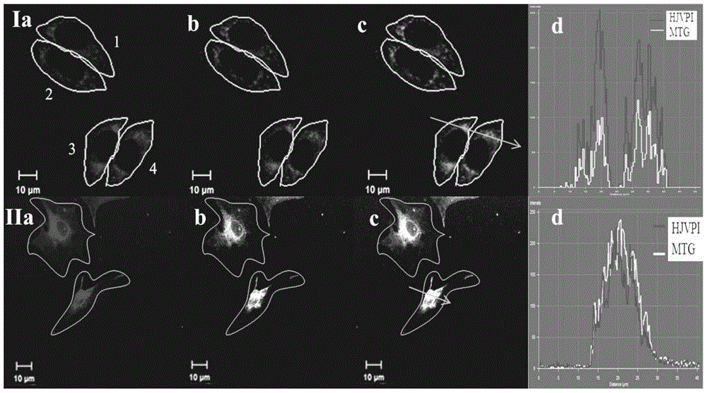
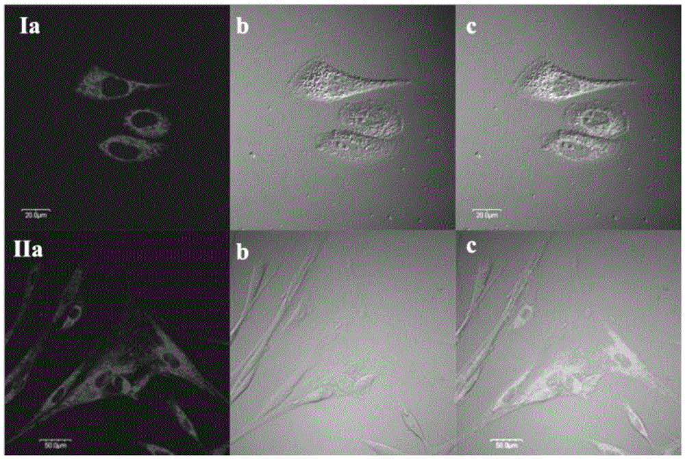
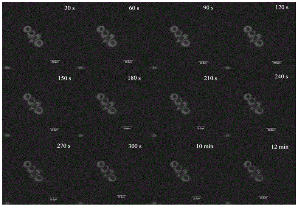
[0030] Example 3: HJVPI staining of SiHa cells / BMSC cells and MTG counterstaining experiments

[0031] Wash the inoculated cell slide three times with PBS, stain the cells with 2 μM HJVPI, and store in CO 2 30min in the incubator. Aspirate the culture medium and wash it three times with PBS to wash off the unbound excess dye, stain the cells with 0.2 μM MTG, and store in CO 2 30min in the incubator. Then take out the stained cell slides, wash off the unbound excess dye solution, cover the cell growth side down on the glass slide, and observe the stained parts, fluorescence distribution and brightness changes of the cells under a laser scanning confocal fluorescence microscope. It was found that MTG and HJVPI had similar intracellular distribution regions, and the calculated average colocalization rate of 4 SiHa cells was 0.91, and the average colocalization rate of 2 BMSC cells was 0.89. Therefore, it is confirmed that the probe HJVPI of the present invention can specifical...


PUM


Abstract

The invention discloses a rotor-type two-photon mitochondrion fluorescence probe. The probe has a chemical name of 4-(2-(8-hydroxy-julolidine) vinyl)-1-picoline iodate, and a chemical structural formula as shown in a formula (I). Experiments prove that the probe is a novel fluorescence probe for selectively marking mitochondria of immortalized living cells (SiHa and HeLa cells) and normal living cells (HUVEC, BMSC and MC3T3-E1 cells). The probe is applicable to a TPM infrared excitation source and multiple laser wavelengths (458nm, 488nm, 514nm, 543nm and 561nm) of LSM. Compared with existing mitochondrion fluorescence probes with similar functions, the probe has the characteristics of low price, good two-photon performance, low excitation energy, good light stability and good biocompatibility, and has a great application value in the field of laser excited fluorescence biomarkers.

Description

technical field [0001] The present invention relates to a molecular rotor-based mitochondrial probe with high signal-to-noise ratio and its application, in particular to a rotor-type double-red mitochondrial fluorescence probe suitable for infrared excitation source of two-photon fluorescence microscope and multiple excitation wavelengths of confocal microscope. Needles and their application in living cells. Background technique [0002] Mitochondria is one of the organelles with important physiological functions in eukaryotic cells, and it is an important place for cells to produce energy through respiration. The shape and number of mitochondria can change, and these changes are often associated with human diseases, such as Parkinson's disease and Alzheimer's disease. Therefore, in situ and real-time clear observation of mitochondria is very necessary for our understanding of related physiological processes and corresponding disease diagnosis and treatment. Currently, flu...

Claims


Application Information

Patent Timeline
Patent Type & Authority Patents(China)
IPC IPC(8): C09K11/06C07D455/04G01N21/64C12Q1/02
Inventor 于晓强张革孙渝明郭丽方
Owner SHANDONG UNIV
Why Eureka
Try Eureka