Image processing method for chest X-ray DR (digital radiography) image rib inhibition

An image processing and image technology, applied in the field of medical informatization, can solve the problems of poor compliance of patients with standardized treatment, small scale of tuberculosis prevention and treatment teams, and complex structure, so as to improve the recognition accuracy and processing efficiency, and make the analysis conclusions more objective and stable. , to avoid the effect of equipment idle and resource waste

Active Publication Date: 2015-12-09
SICHUAN UNIV
View PDF7 Cites 6 Cited by
  • Summary
  • Abstract
  • Description
  • Claims
  • Application Information

AI Technical Summary

Problems solved by technology

[0003] Although China's tuberculosis epidemic situation presents a severe situation of a large number of infected people, a large number of cases, and a large number of existing patients, tuberculosis itself is preventable and treatable; through the promotion of the Chinese government and international assistance, the country provides free tuberculosis diagnosis and treatment; however, Since more than 80% of tuberculosis patients in our country are in rural areas or floating population, the accessibility of medical services is not as good as that of urban residents and non-floating populations, and patients have poor compliance with long-term standard treatment, and often the treatment effect is not good; in view of the serious harm of tuberculosis, The prevention and control work is difficult. The scale of local tuberculosis prevention and control teams at all levels is still small, and the strength and funds cannot meet the needs of prevention and control. It is necessary to strengthen technical and financial investment, establish a combination of medical and prevention mechanisms, and form an effective prevention and control system; currently, in the prevention and control of tuberculosis In terms of implementation, there are two weak and difficult links in early detection and treatment management. First, due to the concealment of tuberculosis incidence and the limitations of detection techniques, the diagnosis and detection of patients mainly relies on the passive detection method that patients have symptoms and go to the hospital for treatment; although through Taking X-rays for physical examination can actively detect pulmonary tuberculosis, but the main group of patients rarely undergo physical examination; this requires research on how to identify high-risk groups and carry out targeted active discovery work; sputum smear staining and microscopic examination are still widely used The method has been used for more than 130 years, and the detection rate is low; secondly, the standard treatment and management dropout rate for diagnosed pulmonary tuberculosis patients is high, especially for multidrug-resistant tuberculosis treatment. A large number of middle-aged patients have been lost, and only a few can persist in treatment; under this situation, the country has gradually launched a screening program for key populations of tuberculosis patients using basic public health services, and provided free anti-tuberculosis treatment to all confirmed patients
[0004] Chest x-ray DR film uses different densities of human tissues to image differences under X-rays to observe lesions in areas with small differences in thickness and density; however, the human tissue structure is complex, and the chest cavity and abdominal cavity contain the main key organs of the human body, including high-density and low-density various organs; therefore, their images overlap each other, and the ribs of the lungs have a particularly large impact on the observation of lung lobes on DR films; if the images of ribs can be weakened or eliminated, subtle lesions can be found more easily; in addition, The objective recording of image data is conducive to the re-examination and comparison of disease diagnosis and treatment; the radiation dose of patients receiving fluoroscopy is also relatively large. It is an indisputable fact that it is harmful; DR film inspection is a double-edged sword for human health; according to the standards established by the International Commission on Radiological Protection according to X-rays, the total risk of radiation is 0.0165 / Sievert, and X-ray chest film In less than half a second, the exposure rate is about 0.045 mSv / s (1 Sievert = 1000 mSv), and the health risks to the crowd are very limited
However, the gonads, eye lenses, breasts and thyroid glands in the human body are particularly sensitive to radiation, and too frequent inspections are not beneficial; suppressing and eliminating the images of lungs and ribs in DR films can take fewer chest images for physical examiners and patients. film, at the cost of lower radiation dose to obtain a doctor's diagnosis of the lesion

Method used


Image

  • Image processing method for chest X-ray DR (digital radiography) image rib inhibition
  • Image processing method for chest X-ray DR (digital radiography) image rib inhibition

Examples


Embodiment Construction

[0036] The present invention will be further described below in conjunction with the accompanying drawings and specific embodiments.

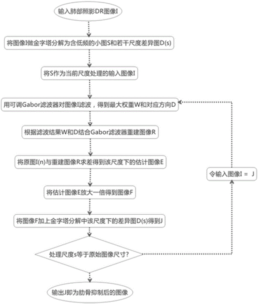
[0037] like figure 1 Shown is an image processing method for chest X-ray DR image rib suppression, comprising the following steps:

[0038] Acquire chest X-ray DR images;

[0039] Decompose the DR image into a pyramid, perform a downsampling process to obtain a Gaussian image pyramid S, and perform an upsampling process to obtain a Laplacian image pyramid difference map D(s);

[0040] Use the smallest S as the current image I to be processed;

[0041] Filter the image I with an adjustable Gabor filter bank to obtain a reconstructed image R;

[0042] Calculate the difference between the image I to be processed and the reconstructed image R, and obtain the processing result image E of the weakened line-segment texture at this scale;

[0043] Double the processing result image E, add it to the corresponding Laplacian image pyramid difference m...


PUM


Abstract

The invention discloses an image processing method for chest X-ray DR (digital radiography) image rib inhibition. The method comprises the following steps: acquiring a chest X-ray DR image; performing pyramid decomposition on the DR image, performing a down sampling process to obtain a Gaussian image pyramid S, and performing an up sampling process to obtain a Laplacian image pyramid difference chart D(S); taking the minimum S as a current to-be-processed image I; performing filtering processing on the image I by using an adjustable Gabor filter bank so as to obtain a reconstructed image R; differencing the to-be-processed image I and the reconstructed image R to obtain a processing result image E with weakened segment-shaped textures under the scale; and doubling the processing result image E, adding the processing result image E and a corresponding Laplacian image pyramid difference chart D(S) under the size together, and repeating the processing procedure until the size is the same as that of an original DR image, thereby obtaining an image after rib inhibition. According to the method disclosed by the invention, the visual saliency of pulmonary shadows is improved, the workload of doctors is reduced, automatic processing can be realized, and an analysis conclusion is more objective and stable.

Description

technical field [0001] The invention relates to the field of medical informatization, in particular to an image processing method for rib suppression in chest X-ray DR images. Background technique [0002] Both DR imaging and traditional chest fluoroscopy use X-ray imaging, which is the main means of physical examination to screen lung diseases, such as lung inflammation, mass, tuberculosis, etc.; however, DR images are digital images with high imaging clarity and low radiation. In practice, it has gradually replaced the traditional chest fluoroscopy imaging. Usually, the purpose of taking DR films is to do tuberculosis screening; tuberculosis is caused by Mycobacterium tuberculosis, which is easily transmitted by droplets or even aerosols in the air; a contagious tuberculosis patient can infect an average of 10 to 15 people in a year; The majority of tuberculosis patients are young and middle-aged, which leads to the loss of family and social labor force; the World Health ...

Claims


Application Information

Patent Timeline
Patent Type & Authority Applications(China)
IPC IPC(8): A61B6/00G06T11/00
Inventor 王俊峰唐鹏高琳姬郁林李虹
Owner SICHUAN UNIV
Why Eureka
Try Eureka