Fusion protein of anthrax toxin receptor cmg2 and human serum albumin

An anthrax toxin receptor, human serum albumin technology, applied in the field of fusion proteins, can solve problems such as uncontrollable

Active Publication Date: 2019-01-18
INST OF BIOENG ACAD OF MILITARY MEDICAL SCI OF THE CHINESE
View PDF1 Cites 0 Cited by
  • Summary
  • Abstract
  • Description
  • Claims
  • Application Information

AI Technical Summary

Problems solved by technology

At the same time, since Fc may also mediate other effects such as ADCC or CDC, these functions are not necessary for prolonging the half-life of receptor-like receptors in vivo, but may cause other uncontrollable effects

Method used


Image

  • Fusion protein of anthrax toxin receptor cmg2 and human serum albumin
  • Fusion protein of anthrax toxin receptor cmg2 and human serum albumin
  • Fusion protein of anthrax toxin receptor cmg2 and human serum albumin

Examples


Embodiment 1

[0029] Embodiment 1: the preparation of fusion protein (CMG2-HSA)

[0030] 1. Expression vector construction

[0031] The fusion protein (CMG2-HSA) expression sequence of CMG2 and albumin was constructed in three steps:

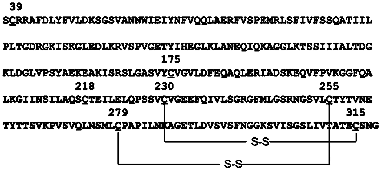
[0032] Step 1: Use primers H-1F and H-1R to amplify the HSA fragment from the plasmid containing the HSA coding sequence cDNA (for example, the T-HSA plasmid kept in our laboratory) by PCR method, and at the same time at its 5' end and An XhoI restriction site and a part of the linker sequence (linker) were added to the 3' end: GGTGGAGGCGGTTCAGGCGG. Use primers CMG2-F and CMG2-R to amplify the sCMG2 fragment from a plasmid containing the sCMG2 coding sequence (for example, PQE30-sCMG2 preserved in our laboratory) by PCR method, and add another part at its 5' end and 3' end, respectively Linker sequence (GTGGCTCTGGCGGTGGCGGATCG) and NotI restriction site. The above steps can also be accomplished by artificially synthesizing nucleotide sequences. Compared w...

Embodiment 2

[0042] Embodiment 2.SPR method detects the affinity of class receptor and PA

[0043] BiacoreT200 (GE) was used and completed at 25°C. Reagents used for coupling, conjugation, and regeneration were all from BIAcore. The reaction buffer is HBS-P+5mM MgCl 2 (10 mM Hepes, 0.15M NaCl, 0.005% P20, pH=7.4). At pH=4.0, 30 μg of PA was immobilized on the CM5 chip by amino coupling. 1 channel as reference channel, only use HBS-P+5mM MgCl 2 as the mobile phase. The regeneration buffer was 10 mM sodium tetraborate, 1 mol / L NaCl, pH=8.5, and all data analyzes were performed using BiacoreT200 evaluation software.

[0044] As shown in Figure 5, CMG2-HSA ( Figure 5A ), sCMG2 ( Figure 5B ) and PA were 3.9nmol / L and 1.915nmol / L respectively. Although CMG2-HSA was slightly lower than sCMG2 in terms of absolute value, it was still at a relatively high affinity level and could reach the nmol / L level.

Embodiment 3

[0045] Example 3. The metabolism of CMG2-HSA in rats

[0046] Two male SD rats (190-210 g, SCXK (Beijing) 2012-0001) were used to inject 100 μg of CMG2-HSA diluted with PBS through the tail vein. Blood was collected at different time points before and after injection until 6 days after injection, and blood was collected 8 times in total. Samples were collected in EP tubes treated with heparin, placed on ice, centrifuged at 4000×g for 15 minutes at 4°C, separated from plasma, and frozen at -20°C until use. The content of CMG2-HSA in the sample was measured by double-antibody sandwich ELISA, and the elimination half-life of CMG2-HSA in vivo was calculated. PBS was coated with 4 μg / mL anti-HSA monoclonal antibody (lifespan biosciences, LS-C51816) on overnight at ℃, wash 4 times with PBST (containing 0.2% Tween-20) the next day, 5 minutes each time; then block with 2% BSA at 37 ℃ for 1 h, repeat the washing step; Add the diluted sample to be tested, incubate at 37°C for 1 h, rep...


PUM

PropertyMeasurementUnit
molecular weightaaaaaaaaaa

Abstract

The invention discloses a fused protein. The first part of the fused protein is recombinant anthrax toxin receptor CMG2, the second part is recombinant human serum albumin, the first part and the second part are connected by a linker peptide, the half-life in vivo of the fused protein is increased by nearly 20 times compared with the recombinant anthrax toxin receptor CMG2. Also, the fused protein has very good in vivo activity, and can fully protect toxin attacked experimental animals.

Description

technical field [0001] The invention relates to a fusion protein and belongs to the technical field of polypeptides. Background technique [0002] Anthrax is a severe zoonotic infectious disease caused by Bacillus anthracis. Because the spores formed by Bacillus anthracis are highly resistant to the external environment, it is a potential biological warfare agent and bioterrorist pathogen. The two main virulence factors of Bacillus anthracis are capsule and exotoxin, among which exotoxin is considered to be the key factor of Bacillus anthracis pathogenicity. Exotoxin consists of three proteins: protective antigen (Protective Antigen, PA), lethal factor (LethalFactor, LF), edema factor (Edema Factor, EF). EF is a calcium ion-dependent adenylate cyclase that can up-regulate intracellular cAMP levels; LF is a zinc-ion-dependent metalloenzyme that can block the MAPKK signaling pathway; the binding of PA to receptors on the cell membrane is LF and EF enter the cell to play a k...

Claims


Application Information

Patent Timeline
Patent Type & Authority Patents(China)
IPC IPC(8): C07K19/00C12N15/62C12N15/81C12N1/19C07K1/22
Inventor 陈薇徐俊杰李亮亮郭强张军董大勇殷瑛付玲
Owner INST OF BIOENG ACAD OF MILITARY MEDICAL SCI OF THE CHINESE
Why Eureka
Try Eureka