
Kit and method for detecting duck hepatitis A viruses through dual TaqMan fluorescent quantitation RT-PCR

A duck hepatitis A virus, fluorescence quantitative technology, applied in microorganism-based methods, biochemical equipment and methods, microorganism determination/inspection and other directions, can solve the problems of no economic loss, difficult to standardize, low sensitivity, etc. Simple, high sensitivity, good specificity

Inactive Publication Date: 2016-01-20
SICHUAN AGRI UNIV
View PDF8 Cites 11 Cited by
  • Summary
  • Abstract
  • Description
  • Claims
  • Application Information

AI Technical Summary

Problems solved by technology

[0003] At present, there are many domestic and foreign reports on the detection methods of DHAV, including serum neutralization test, agar diffusion test and ELISA. Clinically, it often causes huge economic losses due to the inability to make correct diagnosis and effective prevention and control in time.

Method used


Image

  • Kit and method for detecting duck hepatitis A viruses through dual TaqMan fluorescent quantitation RT-PCR
  • Kit and method for detecting duck hepatitis A viruses through dual TaqMan fluorescent quantitation RT-PCR
  • Kit and method for detecting duck hepatitis A viruses through dual TaqMan fluorescent quantitation RT-PCR

Examples


Embodiment 1

[0029] Design and synthesis of embodiment 1 primer and TaqMan probe

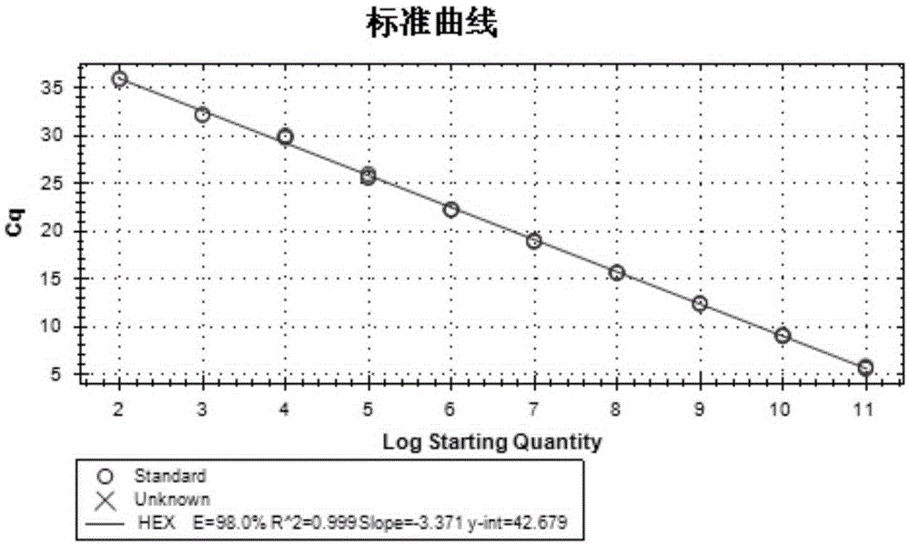
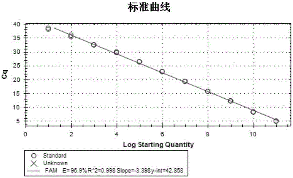
[0030] According to the gene sequences of DHAV-1 (accession number: JQ301467.1), DHAV-2 (accession number: EF067924.1; EF067923.1), DHAV-3 (accession number EU877916.1) in GenBank, by comparison , select the conserved, specific base sequence (nucleotide sequence as shown in SEQIDNO.1, SEQIDNO.2) in the VP0 region of DHAV-1 and the VP3 region of DHAV-3, adopt PrimerPrimer5.0 software to carry out specific primer and For the design of TaqMan probes, see Tables 1 and 2 for details.

[0031] Table 1 Standard primers

[0032]

[0033] Table 2 Quantitative primers and probes

[0034]

[0035] See SEQ ID NO.15 for the expected sequence of PCR amplification of primer pair 1, see SEQ ID NO.16 for the expected sequence of PCR amplification of primer pair 2; see SEQ ID NO.13 for the expected sequence of the quantitative PCR amplification fragment (VP0) of primer pair 3, and see SEQ ID NO. The expected sequence...

Embodiment 2

[0036] Embodiment 2 fluorescence quantitative RT-PCR detects

[0037] 1. Determination of fluorescent quantitative RT-PCR detection method

[0038] 1.1 Sample preparation

[0039] 1.1.1 RNA nucleic acid extraction

[0040] ①Take 0.5mL of allantoic fluid sample into a 1.5mL centrifuge tube treated with DEPC;

[0041] ②Add 0.5mL Trizol, shake and mix well, and let stand at room temperature for 10min;

[0042] ③Add 0.2mL chloroform, vibrate vigorously to mix well, fully emulsify until there is no phase separation, and place at room temperature for 5 minutes;

[0043] ④ Centrifuge at 12000rpm for 15min at 4°C;

[0044] ⑤ Take out the centrifuge tube carefully without shaking, and carefully pipette the supernatant into another 1.5mL centrifuge tube treated with DEPC;

[0045] ⑥ Add an equal volume of isopropanol to the supernatant, invert the centrifuge tube up and down to mix, and let stand at 4°C for 5-10 minutes to precipitate nucleic acids;

[0046] ⑦ Centrifuge at 12000r...

Embodiment 3

[0093] The assembly of embodiment 3 detection kits

[0094] According to the research results of Example 1 and Example 2, a detection kit was assembled for use, and the composition of the kit is shown in Table 10.

[0095] Table 10 Kit Composition

[0096]


PUM


Abstract

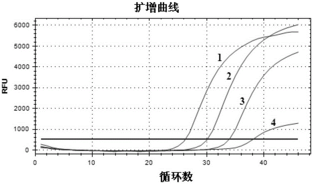
The invention discloses a kit and method for detecting duck hepatitis A viruses through dual TaqMan fluorescent quantitation RT-PCR. Primer pairs and probes applied to the kit comprise primer pairs and probes for detecting type-1 duck hepatitis A virus VP0 genes and type-3 duck hepatitis A virus VP3 genes. After sample RNA is subjected to RT-PCR amplification, the result is automatically analyzed and read through software of an FQ-PCR instrument, and whether the sample contains type-1 duck hepatitis A viruses and type-3 duck hepatitis A viruses or not is judged; if logarithmically-growing specific amplification curves in which Ct<35 exist, it is shown that the sample contains the type-1 duck hepatitis A viruses and / or the type-3 duck hepatitis A viruses; if no logarithmically-growing specific amplification curve in which Ct<35 exists, the sample does not contain the type-1 duck hepatitis A viruses and / or the type-3 duck hepatitis A viruses. The kit and method are adopted for detecting the type-1 duck hepatitis A viruses and the type-3 duck hepatitis A viruses, the two kinds of viruses can be synchronously detected in the same tube, detection time is short, cost is low, the detection result is specific, sensitivity is high, and result judgment is simple and accurate.

Description

technical field [0001] The invention belongs to the technical field of biological detection, and in particular relates to a specific dual fluorescence quantitative RT-PCR (rRT-PCR) kit, primer pair, probe and detection method capable of simultaneously detecting type 1 and type 3 duck hepatitis A viruses. Background technique [0002] Duck viral hepatitis is an important infectious disease that endangers the duck industry, and its main pathogenic factor is picornavirus, namely duck hepatitis A virus (duckhepatitisAvirus, DHAV) of the family Picornaviridae and the genus Avian Hepavirus. According to the whole genome sequence analysis, duck hepatitis A virus can be divided into three independent genotypes, gene type A duck hepatitis A virus (DHAV-A), gene type B duck hepatitis A virus (DHAV-B) and gene type Type C duck hepatitis A virus (DHAV-C); according to serological neutralization experiments, these 3 genotypes have no serum crossover, so they correspond to 3 different ser...

Claims


Application Information

Patent Timeline
IPC IPC(8): C12Q1/70C12Q1/68C12N15/11C12R1/93
CPCC12Q1/6851C12Q2531/113C12Q2537/143C12Q2545/113C12Q2561/101
Inventor 程安春胡琴汪铭书朱德康杨乔贾仁勇陈舜孙昆峰刘马蜂吴英陈孝跃
Owner SICHUAN AGRI UNIV
Learn More