

Surface-enhanced Raman scattering base with visible hot spots, preparation method and method for detecting molecules through base
A surface-enhanced Raman and Raman scattering technology, applied in Raman scattering, material excitation analysis, etc., can solve the problems of uneven distribution of hot spots, large relative average deviation, and inability to meet the requirements of high-sensitivity qualitative analysis and quantitative analysis. achieve the effect of improving sensitivity
- Summary
- Abstract
- Description
- Claims
- Application Information
AI Technical Summary
Problems solved by technology
Method used
Image

Examples
Embodiment 1
[0026] Step 1, preparation of silver nanowires
[0027] Add 10mL of ethylene glycol into a 100mL three-neck flask, heat it to reflux at 160°C for 2h, and inject 0.1M silver nitrate solution and 0.222g of polyvinylpyrrolidone (dissolved in 5mL of ethylene glycol solvent respectively) into it simultaneously with two syringe pumps. Middle), the injection rate was 0.2mL / min. Finally, react at a constant temperature of 160°C for 60 minutes. The synthesized silver nanowire sol is washed twice with acetone and secondary water according to the volume ratio of 1:9. The obtained silver nanowires have a length of 10 μm and a diameter of about 110 nm. .
[0028] Step 2, preparation of gold nanoparticles:
[0029] Add 90mL of secondary water and 1mL of 1% chloroauric acid solution into a 500mL three-neck flask, stir magnetically, heat the oil bath to boiling, then quickly add 1mL of 1% sodium citrate solution as a reducing agent , the diameter of the obtained gold nanoparticles is about...
Embodiment 2
[0035] Use the SERS substrate prepared by the present invention to measure 4-ATP molecules, and obtain SERS spectra and regional SERSmapping images:
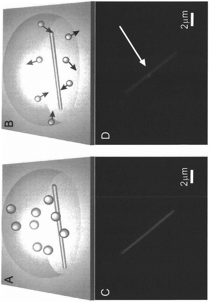
[0036] Place the SERS substrate synthesized in Example 1 under a Raman optical microscope, find the position where the silver nanowire surface is modified with gold nanoparticles, that is, the position where the SERS "hot spot" is generated, and pipette 1 μL with a concentration of 10 -8 M's 4-ATP solution, slowly add dropwise to the "hot spot" or near it. After drying for about 1 minute, select the excitation light with a wavelength of 633nm and a laser power of 2mW, integrate for 1 second, detect the signal, and obtain the SERS spectrum of the 4-ATP molecule.
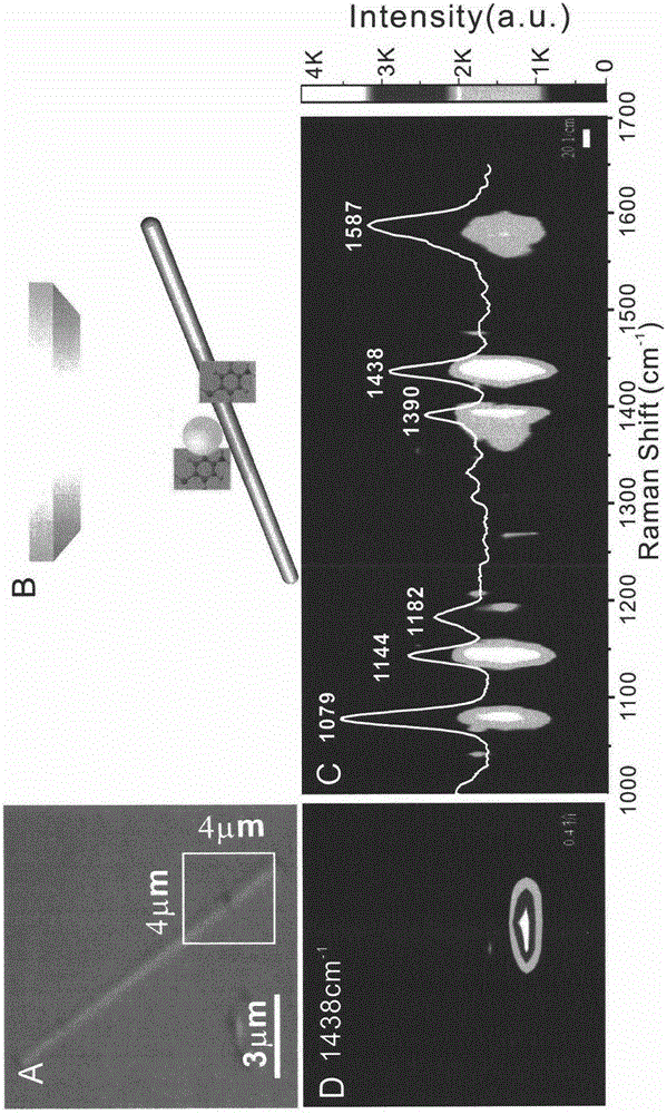
[0037] figure 2 A is the Raman optical microscope image of the SERS substrate prepared by the present invention, where the box marked is the spot range of the Raman laser: (4×4) square microns; figure 2 B is a schematic diagram when the substrate prepared by the pre...
Embodiment 3
[0040] Utilize the SERS substrate prepared by the present invention to measure cysteine, adenosine triphosphate, methamphetamine and paraoxon:
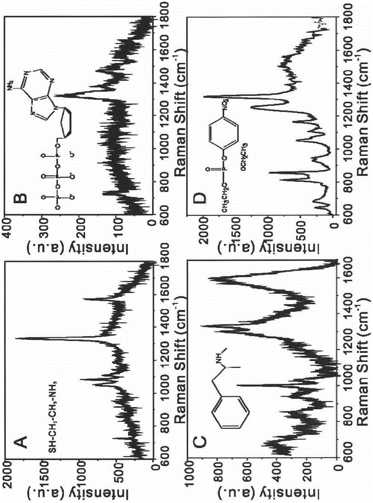
[0041] With the method identical with above-mentioned embodiment 2, can obtain respectively that concentration is 10 -6 M cysteine, 10 -6 M adenosine triphosphate, 10 -7 M of methamphetamine and 10 -8 SERS spectrum of paraoxon in M.
[0042] image 3 Medium, A: 10 -6 Cysteine for M, B: 10 -6 ATP of M, C: 10 -7 Methamphetamine for M, D: 10 -8 M paraoxon, the results show that whether it is common cysteine and adenosine triphosphate molecules in the human body, or common methamphetamine molecules and pesticide paraoxon molecules, the SERS substrate prepared by the invention can achieve high-sensitivity SERS Detection effect.
PUM
| Property | Measurement | Unit |
|---|---|---|
| length | aaaaa | aaaaa |
| diameter | aaaaa | aaaaa |
| length | aaaaa | aaaaa |
Abstract
Description
Claims
Application Information