
Preparation method of slow-release polypeptide growth factor biological material scaffold

A peptide growth factor and biomaterial technology, applied in the field of preparation of slow-release polypeptide growth factor biomaterial scaffolds, can solve the problems of inability to achieve therapeutic effect, short half-life of polypeptide growth factor, influence of polypeptide growth factor biological activity, etc.

Inactive Publication Date: 2016-02-03
BEIHANG UNIV
View PDF3 Cites 4 Cited by
  • Summary
  • Abstract
  • Description
  • Claims
  • Application Information

AI Technical Summary

Problems solved by technology

[0003] Polypeptide growth factors have superior biological activity, but because of the short half-life of polypeptide growth factors in the body, it is not enough to provide nutrition during the recovery of damaged tissues. Therefore, the method of directly injecting polypeptide growth factors in vivo cannot achieve good therapeutic effects. Encapsulate the polypeptide growth factor in the biopolymer carrier. At present, the free polypeptide growth factor is generally encapsulated into the polymer material through a cross-linking agent or in an organic solvent, so that it is in a slow-release state to prolong the life. Its action time
However, in the process of preparing the polypeptide growth factor-biopolymer carrier system, its preparation conditions, physical factors (such as temperature, mechanical stirring intensity, etc.), chemical factors (pH value of solvent, ionic strength of buffer, etc.) The cross-linking agent, etc. will have a certain impact on the biological activity of the polypeptide growth factor

Method used


Image

  • Preparation method of slow-release polypeptide growth factor biological material scaffold
  • Preparation method of slow-release polypeptide growth factor biological material scaffold
  • Preparation method of slow-release polypeptide growth factor biological material scaffold

Examples


Embodiment 1

[0033] Embodiment 1, a kind of preparation method of the chitosan sustained-release scaffold of encapsulating nerve growth factor

[0034] Nerve growth factor (NGF) is one of the members of the neurotrophic factor family and is the first discovered neurotrophic factor, which plays a role in neuronal cell survival, axon regeneration, and regulation of Schwanncell (SC) differentiation and Both play an important role in the process of axonal remyelination. The axons of the central nervous system can regenerate under a suitable microenvironment, and can partially restore the function of damaged axons, but the half-life of nerve growth factor in adult rats is only 7.2min. In this example, nerve growth factor was taken as the test object, and a chitosan slow-release scaffold encapsulated with nerve growth factor was prepared to prolong the slow-release time of nerve growth factor. Specific steps are as follows:

[0035] 1. Chitosan material and nerve growth factor blend swelling ...

Embodiment 2

[0041] Example 2, Bioactivity Verification of Chitosan Slow-release Scaffold Encapsulating Nerve Growth Factor

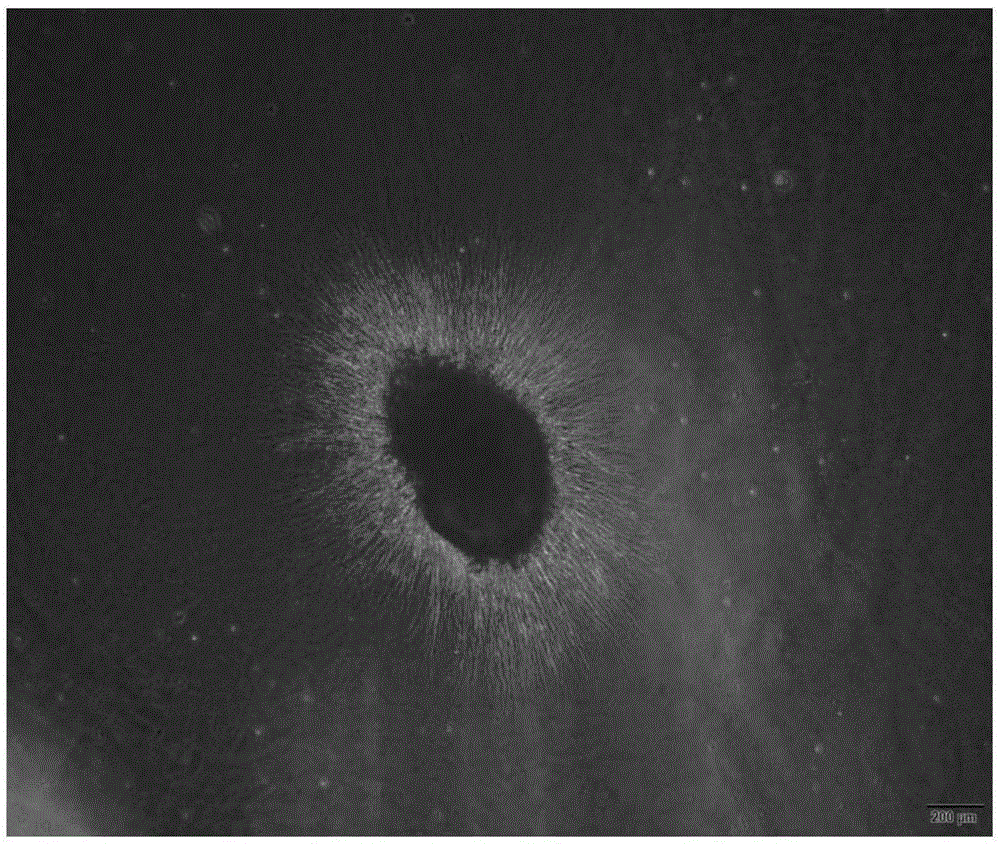
[0042] In order to verify whether the chitosan slow-release scaffold encapsulating nerve growth factor prepared according to the method in Example 1 destroys the activity of nerve growth factor and whether it also has the effect of nourishing neurons, the present invention uses chicken embryo dorsal root nerve The nerve growth factor activity of the chitosan slow-release scaffold encapsulating nerve growth factor obtained in Example 1 was verified by using chicken embryo dorsal root ganglion (DRG) culture method as the test object. Specific steps are as follows:

[0043] Chitosan sustained-release scaffolds encapsulated with nerve growth factor were co-cultured with dorsal root ganglion extracted from 8-day-old chicken embryos. After 48 hours, the growth of DRG axons was observed under an optical microscope.

[0044] The result is as image 3Shown: the dorsal root...

Embodiment 3

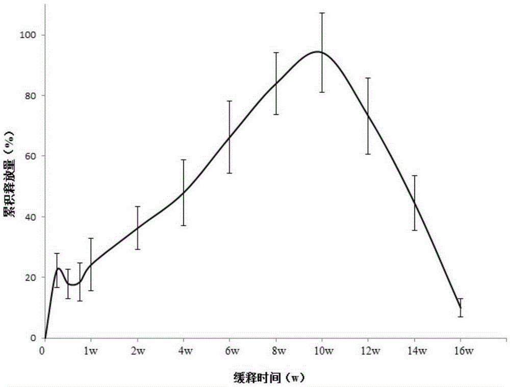
[0045] Embodiment 3, the sustained-release curve of the chitosan sustained-release stent encapsulating nerve growth factor

[0046] After spinal cord injury, the important cause of nerve regeneration impairment after injury is the formation of glial scar. The barrier effect of glial scars on axon regeneration is mainly reflected in two aspects: 1. The mechanical barrier formed by astrocytes; 2. The chemical barrier composed of chondroitin sulfate proteoglycans. The mechanical barrier that hinders axon regeneration begins to enter a stable period 2 months after spinal cord injury, and finally reaches a stable level at 3 months after injury; the chemical barrier that hinders axon regeneration begins to relax 1 month after injury, And reached stability in 3 months. Once the spinal cord injury enters the stage of old spinal cord injury, the stable glial scar and the local environment of the injury tend not to change, and it becomes more difficult to repair the damaged part at thi...


PUM


Abstract

The invention discloses a preparation method of a slow-release polypeptide growth factor biological material scaffold. The preparation method comprises the following steps: (1) uniformly mixing a biological material, a polypeptide growth factor and water for reaction to ensure that the biological material is swollen and the water exists in the biological material in three forms namely combined water, intermediate state water and free water, thereby obtaining a blending system; (2) performing freezing treatment on the blending system to obtain a frozen product; and (3) performing unfreezing treatment on the frozen product to obtain a biological material scaffold releasing the polypeptide growth factor. The slow-release polypeptide growth factor biological material scaffold prepared by the method disclosed by the invention can provide a supporting effect for the polypeptide growth factor to ensure that the polypeptide growth factor is released slowly in a long time, so that while the bioactivity of the polypeptide growth factor is ensured to the utmost extent, a long-time polypeptide growth factor slow release can be provided, thereby providing the polypeptide growth factor required by growth in a whole regeneration process for defective tissues.

Description

technical field [0001] The invention belongs to the field of bioengineering, and in particular relates to a preparation method of a slow-release polypeptide growth factor biomaterial scaffold. Background technique [0002] Polypeptide growth factors are biologically active polypeptides that play an important role in the process of embryogenesis, development, differentiation, and regeneration and repair of various human tissues after injury. The repair of human tissue after injury depends on the nutrition and support of the polypeptide growth factor group. Generally speaking, when the polypeptide growth factor interacts with cells, the growth factor acts as a ligand and binds to the corresponding receptor to form a complex. The complex condenses on the surface of the cell membrane, then vesicles into the cell, and is decomposed due to the presence of lysosomes. The half-life of the polypeptide growth factor in the body is extremely short. [0003] Polypeptide growth factors...

Claims


Application Information

Patent Timeline
IPC IPC(8): A61L27/54A61L27/56A61L27/20A61L27/18A61L27/22
Inventor 李晓光高钰丹杨朝阳段红梅
Owner BEIHANG UNIV
Learn More