A kind of Nile red staining method of poultry primordial germ cells
A technology of primordial germ cells and staining method, which is applied in the field of Nile red staining of poultry primordial germ cells, can solve the problems that cannot be used as a method for cell identification, the staining effect cannot meet the requirements, and the repeatability is not ideal, etc. The effect is stable and repeatable, making up for expensive effects
- Summary
- Abstract
- Description
- Claims
- Application Information
AI Technical Summary
Problems solved by technology
Method used
Image
Examples
Embodiment 1
[0055] Example 1 Obtaining Primordial Germ Cells from Poultry Blood
[0056] (1) Collection of primordial germ cells from blood
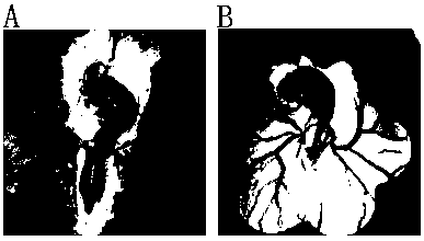
[0057] Hen eggs developed to 55-60 hours and goose eggs developed to 3.5-4 days, the blunt end was opened to expose the embryos, and the embryos were transferred to the PBS solution without calcium and magnesium ions by using a filter paper ring, gently washed, and placed on a plate placed under a microscope (40x magnification), such as figure 1 as shown, figure 1 A is the vascular distribution map of chicken embryos developed to 55-60 h, figure 1 B is the blood vessel distribution map of goose embryos developed to 3.5-4 days. The arrows in the figure show the yolk veins of chicken embryos and goose embryos; use a microneedle to draw blood from the yolk veins on both sides, and finally put it into a container filled with 10% FCS TCM199 centrifuge tubes for use.
[0058] (2) Primordial germ cells were purified by Ficoll 400 density gradient centr...
Embodiment 2
[0061] Example 2 Obtaining Primordial Germ Cells in the Gonads of Poultry
[0062] (1) Separation of gonads
[0063] Chicken embryos develop to 6-7 days, goose embryos develop to 8-9 days, the embryos are taken out from the eggs, the gonads are located above the kidneys, in strip shape, other tissues are carefully peeled off, and washed three times in PBS without calcium and magnesium ions , that is, to obtain the left and right gonads, such as image 3 shown, where image 3 A is the microscope image of chicken embryo gonad, image 3 B is the microscope image of goose embryo gonad.
[0064] (2) Digestion of gonads
[0065] Dilute 0.25% trypsin-EDTA 3 times with PBS without calcium and magnesium ions, then place the left and right gonads in the diluted trypsin-EDTA, digest at 37°C for 5 min to form single cells, and finally add DMEM to stop For digestion, centrifuge at 1500 rpm for 5 minutes, discard the supernatant, and take the precipitate;
[0066] (3) Acquisition of P...
Embodiment 3
[0069] Example 3 PGCs Nile Red staining test
[0070] (1) Dilute the 1mg / mL Nile Red / methanol solution 100 times with PBS without calcium and magnesium, add it to the PGCs cells obtained in Example 1 and Example 2, respectively, blow and beat to form a single cell in suspension, and keep away from room temperature. Light staining for 5-10 min;
[0071] (2) Centrifuge the solution at 1500rpm for 5 minutes, take the cell pellet and add PBS without calcium and magnesium to wash;
[0072] (3) Dilute the Hoechst 33342 solution with PBS without calcium and magnesium to 10 μg / ml, add it to PGCs, and stain in the dark for 2 min;
[0073] (4) Observe the cells under an inverted fluorescent microscope.
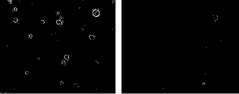
[0074] Microscope (magnification 200 times) detection results are as follows Figure 5-8 Shown are chicken blood PGCs, chicken gonad PGCs, goose blood PGCs and goose gonad PGCs, respectively. Figure 5-8 A in A is the PGCs under bright field, B is the green fluorescence of the PGCs ...
PUM
 Abstract
Description
Claims
Application Information
 - R&D
- Intellectual Property
- Life Sciences
- Materials
- Tech Scout
- Unparalleled Data Quality
- Higher Quality Content
- 60% Fewer Hallucinations
Browse by: Latest US Patents, China's latest patents, Technical Efficacy Thesaurus, Application Domain, Technology Topic, Popular Technical Reports.
© 2025 PatSnap. All rights reserved.Legal|Privacy policy|Modern Slavery Act Transparency Statement|Sitemap|About US| Contact US: help@patsnap.com


