Method for inducing differentiation of embryonic stem cells into liver tissue structure

A technology of embryonic stem cells and liver tissue, applied in the field of cell culture, to achieve the effect of perfect function

Active Publication Date: 2016-05-04
THE FIRST AFFILIATED HOSPITAL OF SUN YAT SEN UNIV
View PDF1 Cites 2 Cited by
  • Summary
  • Abstract
  • Description
  • Claims
  • Application Information

AI Technical Summary

Problems solved by technology

[0006] At present, there is no relevant research to realize the differentiation of embryonic stem cells into real liver tissue structure by enhancing cell adhesion and slowing down the EMT speed in cell differentiation

Method used


Image

  • Method for inducing differentiation of embryonic stem cells into liver tissue structure
  • Method for inducing differentiation of embryonic stem cells into liver tissue structure
  • Method for inducing differentiation of embryonic stem cells into liver tissue structure

Examples


Embodiment 1

[0055] Embodiment 1 Construction of Embryonic Stem Cells Highly Expressing E-cadherin Gene

[0056] (1) Design primers (cDNA-F: 5'-GGAAGCTTATGGGAGCCCGGTGC-3', cDNA-R: 5'-GCGGATCCCTAGTCGTCCTCACCA-3') according to the complete cDNA sequence information of the mouse E-cadherin gene (ACCESSION-AF167441);

[0057] (2) Using the total RNA of the liver tissue of BALB / c mice as a template, reverse transcription to obtain a cDNA template; use the template and the primers involved in step (1) to perform PCR amplification to obtain the E-cadherin gene;

[0058] (3) The PCR product that step (2) obtains is connected with eukaryotic cell pCAG-MCS carrier (purchased from Yrbio company) plasmid connection, constructs pCAG-E-cadherin plasmid, then transforms competent Escherichia coli and cultivates a large amount of extracted plasmids;

[0059] (4) The pCAG-E-cadherin recombinant plasmid was transfected into embryonic stem cells by liposome-mediated method and G418 screening was performed; a...

Embodiment 2 3

[0060] Embodiment 2 three-dimensional culture system establishment

[0061] (2) Establishment of a three-dimensional culture system: collagen scaffold particles (purchased from a reagent company, derived from cow skin collagen), the size of each scaffold particle is about 3×3×2mm, and the internal pore size of the particle is 400um. Each scaffold particle has been micromanipulated Cut to form a 1mm gap to obtain a three-dimensional culture system for semi-open collagen scaffold particles; before use, add culture medium (composed of basal medium and additives; the basal medium is preferably DMEM medium; the additives include: volume fraction of 20%) Fetal bovine serum, 2-mercaptoethanol with a final concentration of 0.1M, HEPES with a final concentration of 25mM, penicillin with a final concentration of 100U / mL, and streptomycin with a final concentration of 100μg / mL) to centrifuge the collagen scaffold particles It was precipitated and then cultured overnight at 37°C.

Embodiment 3

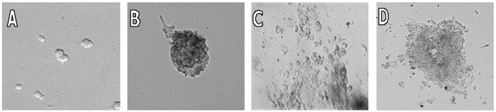
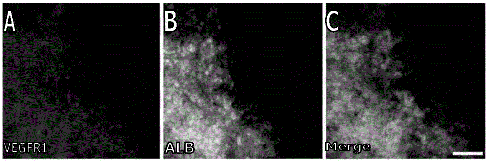
[0062] Example 3 Method for Inducing Differentiation of Embryonic Stem Cells to Liver Tissue Structure

[0063] (1) E-cadherin-ESC cells are induced into embryoid bodies: the E-cadherin-ESC cells prepared in Example 1 were cultured in suspension for 6 days to develop and differentiate into embryoid bodies comprising three germ layer structures; the medium for suspension culture It consists of basal medium and supplements; the basal medium is DMEM medium; the supplements include the following components: fetal calf serum with a volume fraction of 20%, 2-mercaptoethanol with a final concentration of 0.1M , HEPES with a final concentration of 25 mM, penicillin with a final concentration of 100 U / mL and streptomycin with a final concentration of 100 μg / mL;

[0064] (2) Induction of E-cadherin-ESC cells to the liver tissue structure: then transplant the embryoid bodies prepared in step (1) into the three-dimensional culture system of semi-open collagen scaffold particles prepared i...


PUM


Abstract

The invention belongs to the technical field of cell culture and particularly relates to a method for inducing differentiation of embryonic stem cells into the liver tissue structure. E-cadherin genes serve as exogenous genes to transfect the embryonic stem cells, and high-efficiency expression embryonic stem cells of the E-cadherin genes are obtained; then, culture and differentiation are conducted to obtain an embryoid body, the embryoid body is transplanted into a three-dimensional cell culture system containing an EMT inhibitor for induced culture, and the three-dimensional growing liver tissue structure is obtained. According to the method, ESC in-vitro liver tissue structure differentiation is researched in the perspectives of cell adhesion and the EMT level for the first time, the expression level is stably controlled after E-cadherin transfection, and enough cell adhesion can be kept in cell differentiation, and the cells can not be dispersed into monolayer cells easily. The excessively quick EMT level of the embryonic stem cells in-vitro differentiation is reduced, synchronous differentiation of differentiated parenchymal liver cells and hepatic interstitial cells is controlled to form liver tissue, and the method meets the requirement of an in-vivo tissue development mechanism better.

Description

technical field [0001] The invention belongs to the technical field of cell culture, and in particular relates to a method for inducing embryonic stem cells to differentiate into liver tissue structures. Background technique [0002] In the current study on the differentiation of embryonic stem cells (ESC) towards the liver, the level of differentiation research is still mostly limited to the single cell state, that is, it can only differentiate into liver lineage cells such as hepatocytes and bile duct epithelial cells (BEC). The disadvantages include a single cell type and the inability to Further form the liver tissue structure. The existing technology has also successfully induced embryonic stem cells to differentiate into liver cells and bile duct epithelial cells, but the differentiated cells are still in a single cell state and it is difficult to form liver tissue. Some scholars have studied to improve the state of hepatocytes differentiated from embryonic stem cells...

Claims


Application Information

Patent Timeline
Patent Type & Authority Applications(China)
IPC IPC(8): C12N5/10C12N5/071
CPCC12N5/067C12N2500/24C12N2500/44C12N2500/84C12N2501/11C12N2501/12C12N2501/148C12N2501/15C12N2501/237C12N2501/33C12N2506/02C12N2510/00C12N2513/00
Inventor 胡安斌
Owner THE FIRST AFFILIATED HOSPITAL OF SUN YAT SEN UNIV
Why Eureka
Try Eureka