
Super-resolution optical imaging probe for living cells and preparation method of super-resolution optical imaging probe

An optical imaging and super-resolution technology, applied in chemical instruments and methods, material analysis by optical means, scientific instruments, etc., can solve the problems of poor photostability, insufficient quantum yield, poor targeting specificity, etc. To achieve the effect of simple and convenient operation, saving time cost and economic cost, and low cost

Active Publication Date: 2016-09-21
SOUTHEAST UNIV
View PDF4 Cites 5 Cited by
  • Summary
  • Abstract
  • Description
  • Claims
  • Application Information

AI Technical Summary

Problems solved by technology

However, in terms of the characteristics of the probes required by the above-mentioned PALM / STORM technology, these existing optically labeled probes have more or less shortcomings, such as insufficient quantum yield, poor photostability, and poor targeting specificity. Wait

Method used


Image

  • Super-resolution optical imaging probe for living cells and preparation method of super-resolution optical imaging probe
  • Super-resolution optical imaging probe for living cells and preparation method of super-resolution optical imaging probe

Examples


Embodiment Construction

[0017] Below in conjunction with embodiment the present invention will be further described.

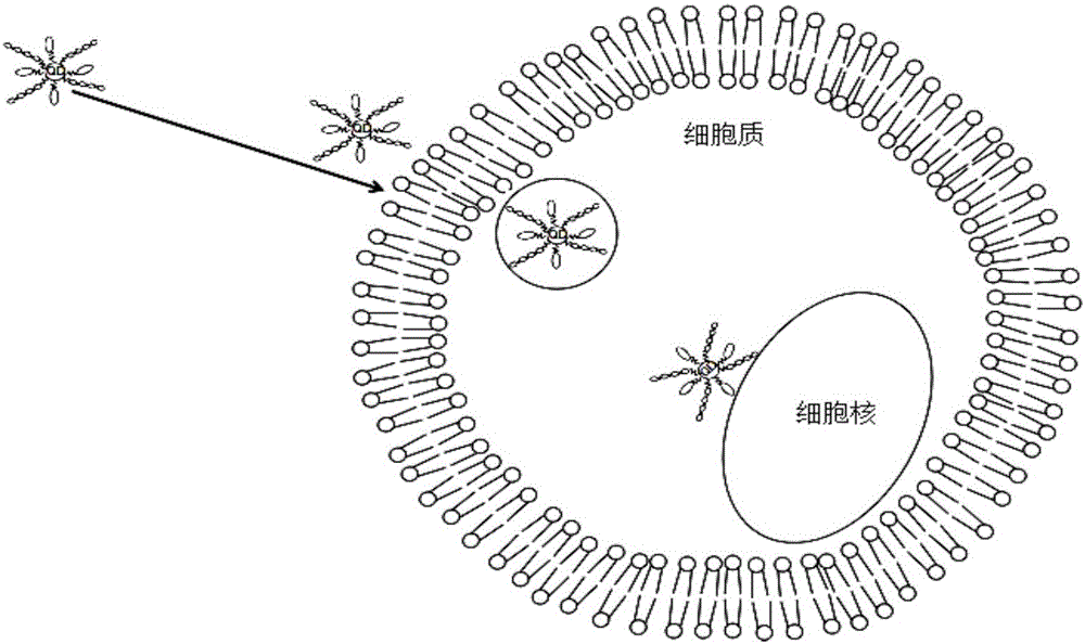
[0018] The quantum dots involved in this example are CdSe / ZnS oil-phase quantum dots, and their fluorescence emission wavelength is 640nm; the carboxyl small molecule ligand involved is thioglycolic acid; the cell membrane penetrating peptide involved is YGRKKRRQRRR, and the nucleic acid aptamer involved The sequence is 5'-NH 2 - TTGGTGGTGGTGGTTGTGGTGGTGGTGG-3'; the cells involved are HeLa cells.

[0019] When preparing the probe, first take 500 μL of CdSe / ZnS quantum dots, add 1 mL of methanol and centrifuge at 3000 rpm for 10 min, discard the supernatant and disperse the precipitate into 2 mL of chloroform. After adding 100 μL tetramethylammonium hydroxide and 100 μL thioglycolic acid, stir vigorously for 2 h. Add 2 mL of deionized water, and collect the quantum dots modified with carboxyl ligands in the upper layer after static layering.

[0020] After adding 4 mg of 1-(3-dimet...


PUM


Abstract

The invention discloses a super-resolution optical imaging probe for living cells and a preparation method of the super-resolution optical imaging probe. Quantum dots are taken as a fluorophore, cell membrane penetrating peptides and nucleic acid aptamers are coupled on the surfaces of the quantum dots, the cell membrane penetrating peptides are used for promoting the probe to penetrate through the living cells, the nucleic acid aptamers are used for specific binding with nuclear membrane receptor protein of the living cells, and cell nucleus positioning of the probe is realized. The quantum dots with scintillating effects are taken as the fluorophore and are adapted to super-resolution optical imaging based on a single-molecule positioning method by means of the high luminous intensity and the fluorescence scintillation characteristic; meanwhile, the quantum dots with scintillating effects are taken as the fluorophore, a special imaging buffering solution is not required, therefore, the probe is adapted to super-resolution imaging of the living cells; besides, the preparation method is simple and convenient to operate, the cost is low, and the time cost and the economic cost are effectively saved.

Description

technical field [0001] The invention relates to a living cell super-resolution optical imaging probe and a preparation method thereof, which belong to nanometer material technology. Background technique [0002] The resolution of optical microscopy is limited by the diffraction limit, usually only to 200-300nm. In recent years, scientists have developed several types of super-resolution optical microscopes that can break through the diffraction limit, such as stimulated emission depletion microscopy (STED), structured light illumination microscopy (SIM), and stochastic optical reconstruction microscopy. (STORM) and photoactivated localization microscopy (PALM). Among them, the resolution achieved by STORM / PALM technology is the most prominent, and their imaging principles are similar, both based on single-molecule localization technology. The 2014 Nobel Prize in Chemistry was awarded to three scientists who made groundbreaking achievements in the field of super-resolution ...

Claims


Application Information

Patent Timeline
IPC IPC(8): C09K11/02C09K11/88G01N21/64
CPCC09K11/025C09K11/883G01N21/6486
Inventor 宗慎飞蒋晓月王著元崔一平
Owner SOUTHEAST UNIV
Learn More