Bovine serum albumin coated ferriferrous oxide nano-particle T1-MRI (Magnetic Resonance Imaging) contrast medium and preparation method thereof

A technology of bovine serum albumin and ferric tetroxide, which is applied in the field of nanomaterials, can solve the problems of absorption offset and changes in the properties of nanomaterials, and achieves the effects of low toxicity, good biocompatibility and broad application prospects.

Inactive Publication Date: 2016-11-09
SHANGHAI NORMAL UNIVERSITY
View PDF4 Cites 15 Cited by
  • Summary
  • Abstract
  • Description
  • Claims
  • Application Information

AI Technical Summary

Problems solved by technology

But this two-step method is prone to lead to changes in the properties of the nanomaterial before and after the exchange, for example, the absorption will shift.

Method used


Image

  • Bovine serum albumin coated ferriferrous oxide nano-particle T1-MRI (Magnetic Resonance Imaging) contrast medium and preparation method thereof
  • Bovine serum albumin coated ferriferrous oxide nano-particle T1-MRI (Magnetic Resonance Imaging) contrast medium and preparation method thereof
  • Bovine serum albumin coated ferriferrous oxide nano-particle T1-MRI (Magnetic Resonance Imaging) contrast medium and preparation method thereof

Examples


Embodiment 1

[0030]①Weigh 0.0675g of BSA into a three-necked flask, add 25mL of secondary water and ultrasonically dissolve it;

[0031] ②Then stir while heating, and nitrogen gas is removed in the flask to remove oxygen, and iron source solution is added thereto (iron source solution consists of 0.0692g FeCl 3 ·6H 2 O and 0.0351 g FeSO 4 ·7H 2 O is miscible in 1mL of secondary water to make);

[0032] ③ After 10 minutes, change to a nitrogen balloon for protection, and use 2 mL of ethanol to eliminate the foam generated by nitrogen gas, so that the solution is completely clarified.

[0033] ④ When the temperature rises to 75°C, quickly add 2.5mL of 28% concentrated ammonia water, keep the reaction at 75°C for 15 minutes, and then the reaction ends.

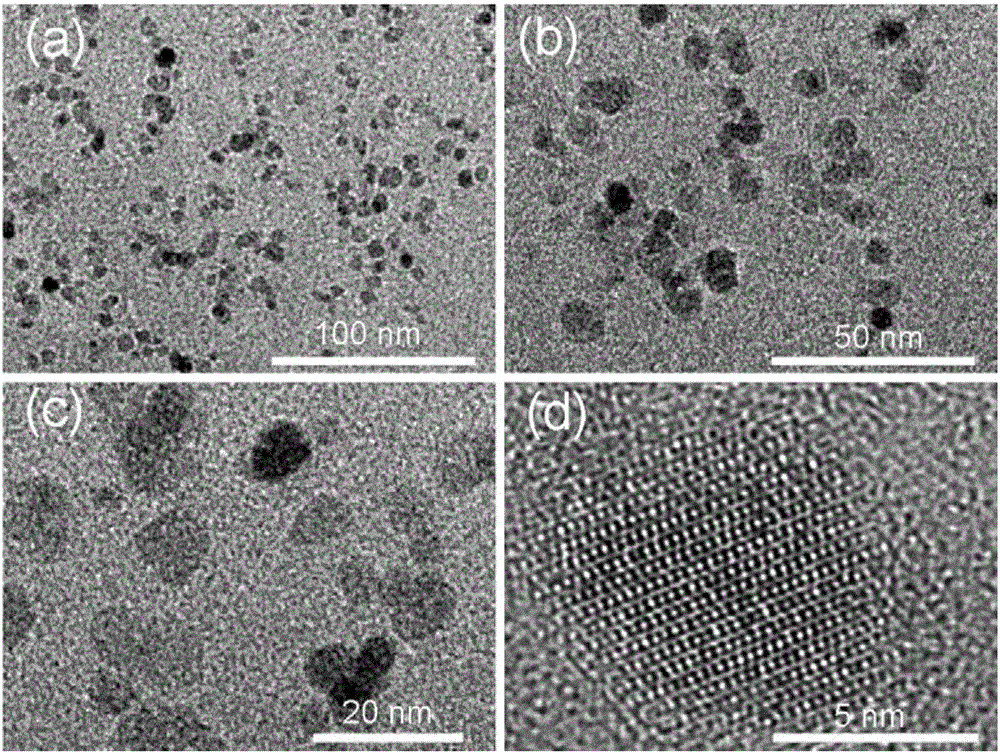
[0034] like figure 1 Shown, for the obtained BSA@Fe prepared in this example 3 o 4 TEM images of different resolutions, from the analysis of the images, it can be known that the prepared BSA@Fe 3 o 4 Particles have good dispersion an...

Embodiment 2

[0041] ①Weigh 0.0833g of BSA into a three-neck flask, add 25mL of secondary water and ultrasonically dissolve it;

[0042] ②Then stir while heating, and nitrogen gas is removed in the flask to remove oxygen, and iron source solution is added thereto (iron source solution consists of 0.0692g FeCl 3 ·6H 2 O and 0.0351 g FeSO 4 ·7H 2 O is miscible in 1mL of secondary water to make);

[0043] ③ After 10 minutes, change to a nitrogen balloon for protection, and use 2 mL of ethanol to eliminate the foam generated by nitrogen gas, so that the solution is completely clarified.

[0044] ④ When the temperature rises to 75°C, quickly add 2.5mL of 28% concentrated ammonia water, keep the reaction at 75°C for 15 minutes, and then the reaction ends.

Embodiment 3

[0046] ①Weigh 0.125g of BSA into a three-necked flask, add 25mL of secondary water and ultrasonically dissolve it;

[0047] ②Then stir while heating, and nitrogen gas is removed in the flask to remove oxygen, and iron source solution is added thereto (iron source solution consists of 0.0692g FeCl 3 ·6H 2 O and 0.0351 g FeSO 4 ·7H 2 O is miscible in 1mL of secondary water to make);

[0048] ③ After 10 minutes, change to a nitrogen balloon for protection, and use 2 mL of ethanol to eliminate the foam generated by nitrogen gas, so that the solution is completely clarified.

[0049] ④ When the temperature rises to 75°C, quickly add 2.5mL of 28% concentrated ammonia water, keep the reaction at 75°C for 15 minutes, and then the reaction ends.


PUM


Abstract

The invention relates to a bovine serum albumin coated ferriferrous oxide nano-particle T1-MRI (Magnetic Resonance Imaging) contrast medium and a preparation method thereof. The method specifically comprises the following steps: (1) dissolving bovine serum albumin into secondary water A; ultrasonically dissolving to prepare a bovine serum albumin solution; (2) adding soluble ferric iron salts and soluble ferrous salts into secondary water B and dissolving to prepare an iron source solution; (3) putting the bovine serum albumin solution into an inert atmosphere and heating and stirring; adding the iron source solution into the bovine serum albumin solution to prepare a BSA-Fe precursor; and (4) raising the temperature to 50-100 DEG C and rapidly adding concentrated ammonia water, and reacting at a constant temperature for 5min-30min to prepare the bovine serum albumin coated ferriferrous oxide nano-particle T1-MRI contrast medium. Compared with the prior art, the preparation method is simple and the controllability is good; and obtained bovine serum albumin coated ferriferrous oxide nano-particles have the characteristics of good biocompatibility, easiness of being discharged out of a body, small toxicity and good T1 imaging effect.

Description

technical field [0001] The invention belongs to the technical field of nanomaterials, and relates to an ultra-small bovine serum albumin-coated ferric oxide nanoparticle T 1 -MRI contrast agent and method for its preparation. Background technique [0002] Malignant tumors have the characteristics of high mortality, difficult treatment and rapid deterioration, and have always been the number one killer of human life. Therefore, early diagnosis and specific treatment of tumors are particularly important. Currently, tumor detection methods mainly include ultrasound imaging, CT imaging, nuclear medicine PET imaging, and magnetic resonance imaging (MRI). With the development of nuclear magnetic resonance technology, the scanning time is gradually shortened, the resolution is gradually improved, and the detection of small lesions is more accurate, which also makes nuclear magnetic resonance imaging technology a new disease detection method developed in recent years. In order to...

Claims


Application Information

Patent Timeline
Patent Type & Authority Applications(China)
IPC IPC(8): A61K49/14A61K49/18
CPCA61K49/143A61K49/1869
Inventor 田启威王媛媛杨仕平安璐芮西川
Owner SHANGHAI NORMAL UNIVERSITY
Why Eureka
Try Eureka