Calcium magnesium silicate porous ceramic ball ocularprosthesis seat and preparation method thereof

A technology of porous ceramic balls and magnesium silicate, applied in the field of medical biomaterials, can solve the problems of slow vascularization efficiency, exposure of prosthetic eye seat, limited biological activity, etc., to achieve the effect of facilitating cell migration and reducing complex problems

Active Publication Date: 2017-02-01
ZHEJIANG UNIV
View PDF6 Cites 11 Cited by
  • Summary
  • Abstract
  • Description
  • Claims
  • Application Information

AI Technical Summary

Problems solved by technology

However, exposure of the HA prosthetic socket may occur once vascularization is delayed
In addition, β-tricalcium phosphate ceramic porous prosthetic eye sockets have also entered the market and entered the clinic. Although this material is better than HA in degradability, it still faces problems such as limited biological activity and slow vascularization efficiency.

Method used


Image

  • Calcium magnesium silicate porous ceramic ball ocularprosthesis seat and preparation method thereof
  • Calcium magnesium silicate porous ceramic ball ocularprosthesis seat and preparation method thereof
  • Calcium magnesium silicate porous ceramic ball ocularprosthesis seat and preparation method thereof

Examples


Embodiment 1

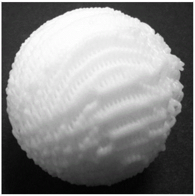
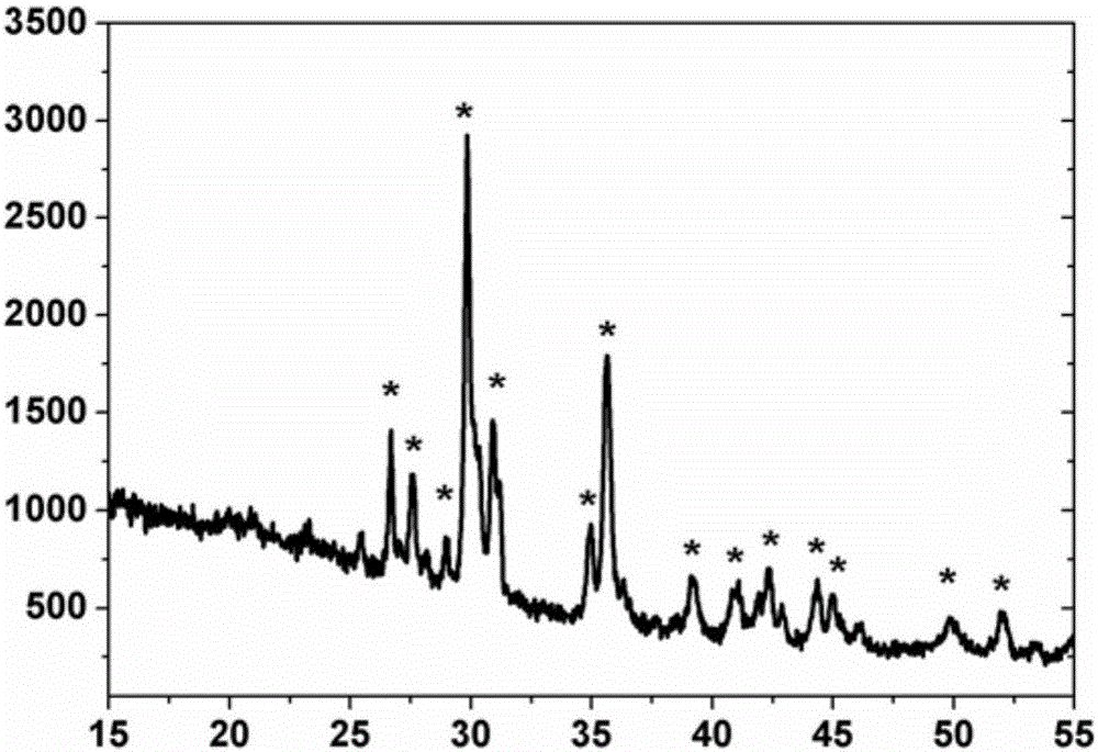
[0044] 1) Put the diopside powder into a ball mill and mill it for 4 hours, sieve with a sieve to obtain an ultrafine diopside powder below 60 μm, and then stir and disperse the ultrafine powder in a solid-to-liquid mass ratio of 1:2 In the polyvinyl alcohol aqueous solution with a concentration of 8%, a two-phase uniform paste is formed, and then the paste is placed in the injection reservoir of the three-dimensional printer connected to the tubular nozzle, and then the three-dimensional printer is started, according to the preset square channel and The diameter is three-dimensionally printed to form a spherical porous composite material with a diameter of 24mm, and then the porous composite material is dried at 60°C for 12 hours and sintered at 1350°C for 8 hours to obtain diopside porous ceramic balls. After testing, sintering The diameter of the final ceramic ball is 22mm, and the channel size is 800μm. figure 1 The appearance shown in the XRD test proves that the sintered...

Embodiment 2

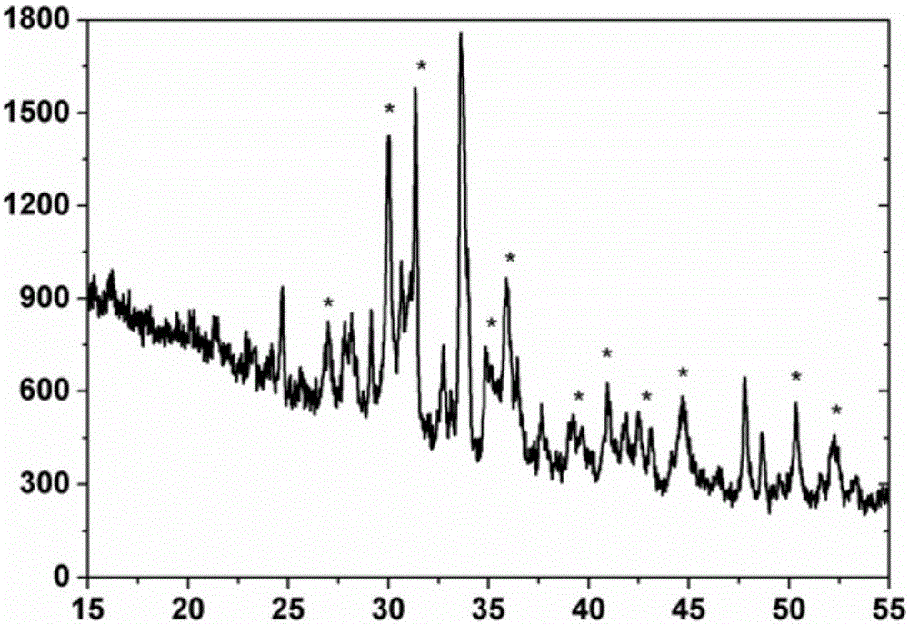
[0048] The preparation method is the same as that in Example 1, the difference is that the composition of the raw materials for preparing the calcium-magnesium silicate modified layer in step 2) is adjusted to: dissolve 0.40 mol of tetraethyl orthosilicate in 400 ml of deionized water, then add 8 ml of nitric acid and stir After 90 minutes, add 0.7mol calcium nitrate and 0.1mol magnesium nitrate, stir and hydrolyze for 90 minutes at room temperature, age for 12 hours, the mixed solution is converted into a hydrogel, and the diopside porous ceramic ball prepared in step 1) is placed in the In the gel, impregnate with negative pressure suction, take out the porous ceramic ball after standing for 8 minutes, then use vacuum filtration for 2 minutes to suck out the excess gel in the pores, continue to age for 36 hours, dry at 95°C for 24 hours, and then calcined at 1200°C for 1 Hours, cool down naturally. After testing, the surface of the diopside porous ceramic ball pore wall has ...

Embodiment 3

[0051] The preparation method is the same as in Example 1, the difference is that the composition of the raw materials for preparing the calcium-magnesium silicate modified layer in step 2) is adjusted to: dissolve 0.20 mol of tetraethylorthosilicate in 90 ml of deionized water, then add 3 ml of nitric acid and stir After 40 minutes, add 0.3mol calcium nitrate and 0.1mol magnesium nitrate, stir and hydrolyze for 60 minutes at room temperature, age for 2 hours, the mixed solution is converted into a gel, and the diopside porous ceramic ball prepared in step 1) is placed in the gel. In the glue, negative pressure suction impregnation, after standing for 5 minutes, take out the porous ceramic ball, then use vacuum filtration for 5 minutes to suck out the excess gel in the pores, continue to age for 36 hours, dry at 95°C for 36 hours, and then calcinate at 1000°C for 2 hours , natural cooling. After testing, the surface of diopside porous ceramic ball channel wall layered with a t...


PUM

PropertyMeasurementUnit
diameteraaaaaaaaaa
thicknessaaaaaaaaaa
thicknessaaaaaaaaaa

Abstract

The present invention discloses a calcium magnesium silicate porous ceramic ball ocularprosthesis seat and a preparation method. The calcium magnesium silicate porous ceramic ball ocularprosthesis seat comprises a non-biodegradable calcium magnesium silicate porous ceramic ball ocularprosthesis seat and a degradable calcium magnesium silicate modification layer covering the surface of the pore channel wall, wherein the ocularprosthesis seat has a completely penetrating porous structure, the porosity is 35-85%, the pore channel diameter is 60-800 [mu]m, the ocularprosthesis seat is a porous diopside ceramic ball constructed by using a three-dimensional printing technology, and the pore channel wall is subjected to degradable calcium magnesium silicate gel precursor filling modification and secondary sintering to obtain the product. According to the present invention, after the calcium magnesium silicate porous ceramic ball ocularprosthesis seat is implanted into the eye socket, the neovessel growth is promoted through the bioactivity of the pore channel surface layer calcium magnesium silicate layer so as to achieve the rapid vascularization in the pore channel and avoid the displacement or prolapse of the ocularprosthesis seat; and the calcium-silicon-based ceramic ocularprosthesis seat pore channel wall bioactivity is excellent, and the application value is provided in the reconstruction of the ocular base.

Description

technical field [0001] The invention relates to medical biomaterials, in particular to a calcium-magnesium silicate porous ceramic spherical artificial eye socket and a preparation method thereof. [0002] technical background [0003] Enucleation is the preferred solution for the treatment of severe rupture of the eyeball, glaucoma, and intraocular tumors. However, the collapse of the upper eyelid, inability to open and close the eyelid, and sunken eye sockets caused by the loss of the eyeball after surgery will have subsequent impacts on the patient. Prosthetic eye seat implantation surgery to compensate for the loss of orbital content is an ideal solution for repairing the loss of the eyeball. The currently clinically used prosthetic eye sockets are mainly solid spheres represented by silica gel and porous prosthetic eye sockets represented by coral phase inversion hydroxyapatite (HA) (US Patent. No. 4976731). After the former is implanted in the eye socket, a fibrous mem...

Claims


Application Information

Patent Timeline
Patent Type & Authority Applications(China)
IPC IPC(8): A61L27/56A61L27/02A61L27/50A61L27/10
Inventor 苟中入何冬霜柳丽敏贺永邵慧锋杨贤燕
Owner ZHEJIANG UNIV
Why Eureka
Try Eureka