Method for detecting blood fluke DNA

A technology of schistosomiasis and amplification system, which is applied in the direction of biochemical equipment and methods, microbial measurement/inspection, etc. It can solve the problems of false positives, long time consumption, contamination of amplification products, etc., and achieve the effect of simple operation and short time

Inactive Publication Date: 2017-03-08
JIANGSU INST OF PARASITIC DISEASES +1
View PDF5 Cites 2 Cited by
  • Summary
  • Abstract
  • Description
  • Claims
  • Application Information

AI Technical Summary

Problems solved by technology

The PCR and Q-PCR methods rely on thermal cycle instruments, have high requirements on the operating environment and personnel, and take a long time. The problem with the LAMP method is that the amplification products will pollute the en

Method used


Image

  • Method for detecting blood fluke DNA
  • Method for detecting blood fluke DNA
  • Method for detecting blood fluke DNA

Examples


Embodiment 1

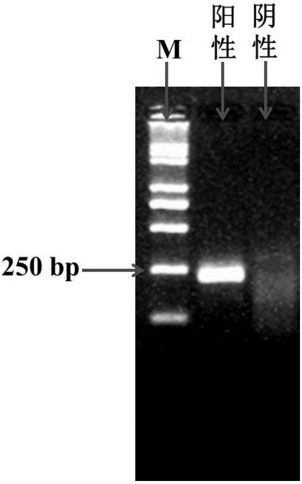
[0060] Sample source: Genomic DNA extracted from adults of Schistosoma japonicum. The concentration of DNA is 50ng / μL.

[0061] The following primers for schistosome were designed and commissioned by Shenggong Bioengineering (Shanghai) Co., Ltd. to synthesize:

[0062] Forward primer sequence: 5’-TACCTCAAGAAGTAATGTCCTTCCATTGTG-3’,

[0063] Reverse primer sequence: 5'-ATGCGAGGTTTCAGGAGACCAAGAAGAACG-3'.

[0064] Preparation of amplification system

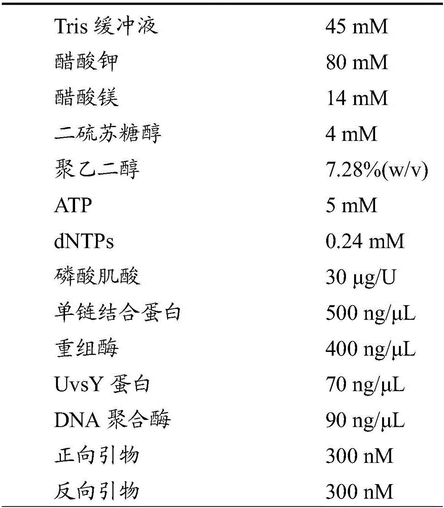
[0065] Prepare the isothermal nucleic acid amplification system in a 200μL centrifuge tube according to the following ratio (the volume is 50μL):

[0066]

[0067] The amplification system prepared above is freeze-dried under negative pressure in a freeze dryer to become a powdery amplification system.

[0068] Add polyethylene glycol with a final concentration of 6% (w / v) and a molecular weight of 35000 as the reaction buffer to the centrifuge tube to re-dissolve the amplification system to 49 μL, and then add 1 μL of the prepared schistosome g...

Embodiment 2

[0071] Sample source: Genomic DNA extracted from adults of Schistosoma japonicum. The concentration of DNA was 70ng / μL.

[0072] The following primers for schistosome were designed and commissioned by Shenggong Bioengineering (Shanghai) Co., Ltd. to synthesize:

[0073] Forward primer sequence: 5’-TACCTCAAGAAGTAATGTCCTTCCATTGTG-3’,

[0074] Reverse primer sequence: 5'-ATGCGAGGTTTCAGGAGACCAAGAAGAACG-3'.

[0075] Preparation of amplification system

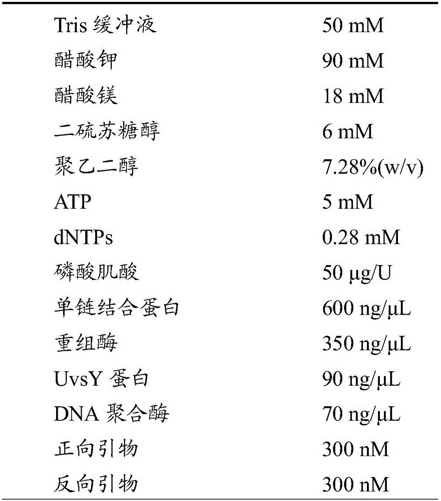
[0076] Prepare the isothermal nucleic acid amplification system in a 200μL centrifuge tube according to the following ratio (the volume is 100μL):

[0077]

[0078] The amplification system prepared above is freeze-dried under negative pressure in a freeze dryer to become a powdery amplification system.

[0079] Add polyethylene glycol with a final concentration of 6% (w / v) and a molecular weight of 35000 as the reaction buffer to the centrifuge tube to redissolve the amplification system to 98 μL, then add 2 μL of the prepared schistosome geno...

Embodiment 3

[0081] Sample source: Genomic DNA extracted from adults of Schistosoma japonicum, the concentration of DNA was 82ng / μL.

[0082] The following primers for schistosome were designed and commissioned by Shenggong Bioengineering (Shanghai) Co., Ltd. to synthesize:

[0083] Forward primer sequence: 5’-TACCTCAAGAAGTAATGTCCTTCCATTGTG-3’,

[0084] Reverse primer sequence: 5'-ATGCGAGGTTTCAGGAGACCAAGAAGAACG-3'.

[0085] Preparation of amplification system

[0086] Prepare the isothermal nucleic acid amplification system in a 200μL centrifuge tube according to the following ratio (the volume is 50μL):

[0087]

[0088] The amplification system prepared above is freeze-dried under negative pressure in a freeze dryer to become a powdery amplification system.

[0089] Add polyethylene glycol with a final concentration of 6% (w / v) and a molecular weight of 35000 as the reaction buffer to the centrifuge tube to re-dissolve the amplification system to 49 μL, and then add 1 μL of the prepared schistosome ...


PUM


Abstract

The invention provides a method for detecting blood fluke DNA. The method comprises the steps that an amplification system and the blood fluke DNA are mixed, isothermal amplification is conducted for 5-40 min at the temperature of 35 DEG C to 45 DEG C to obtain an amplification product, and agarose gel electrophoresis is conducted on the amplification product to detect the blood fluke DNA, wherein the amplification system is prepared from a Tris buffer solution, potassium acetate, magnesium acetate, dithiothreitol, polyethylene glycol, ATP, dNTPs, phosphocreatine, single-strand binding protein, recombinase, UvsY protein, DNA polymerase and specific primers of the blood fluke DNA. The method does not depend on a thermal cycler and is easy to operate, the blood fluke DNA can be detected at the low temperature (from 35 DEG C to 45 DEG C), the amplification time is short (5-40 min), and therefore the method is suitable for basic and site detection.

Description

Technical field [0001] The invention relates to the field of molecular biology detection, in particular to a method for detecting schistosoma DNA. Background technique [0002] Schistosomiasis japonicum is a worldwide zoonotic parasitic disease. It is one of the "six tropical diseases" defined by the World Health Organization (WHO) and one of the five major parasitic diseases in my country. Schistosomiasis is caused by cercariae infecting end hosts such as humans and animals in contact with infected water. The larvae develop in the final host body by absorbing the host nutrition, causing the host malnutrition; through the transformation of antigens, antigen camouflage and weakening the host's immunity, it resists the killing effect of host immunity, and gradually develops into adults, and parasitizes in the portal-mesenchymal vein. system. Symptoms in the acute phase include fever, hepatomegaly, abdominal pain, diarrhea, and blood in the stool; symptoms in the chronic phase are...

Claims


Application Information

Patent Timeline
IPC IPC(8): C12Q1/68
CPCC12Q1/6844C12Q2531/119C12Q2522/101C12Q2527/125
Inventor 杨坤赵松李伟张键锋羊海涛郭利川刘小曼王智宏应清界
Owner JIANGSU INST OF PARASITIC DISEASES
Why Eureka
Try Eureka