
Method for double-signal in-situ detection of intracellular microRNA based on gold-upconversion nanoparticle tetrahedron

A nanoparticle, in situ detection technology, applied in the determination/inspection of microorganisms, biochemical equipment and methods, etc., can solve the problem of cancer cells to be explored and so on

Inactive Publication Date: 2017-03-15
JIANGNAN UNIV
View PDF6 Cites 13 Cited by
  • Summary
  • Abstract
  • Description
  • Claims
  • Application Information

AI Technical Summary

Problems solved by technology

However, the application in living cancer cells in the biological field remains to be explored

Method used


Image

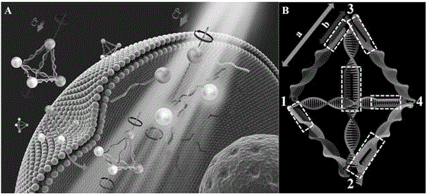
  • Method for double-signal in-situ detection of intracellular microRNA based on gold-upconversion nanoparticle tetrahedron
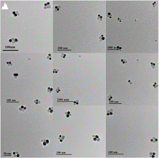
  • Method for double-signal in-situ detection of intracellular microRNA based on gold-upconversion nanoparticle tetrahedron
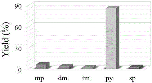
  • Method for double-signal in-situ detection of intracellular microRNA based on gold-upconversion nanoparticle tetrahedron

Examples


Embodiment 1

[0037] Example 1 Gold nanoparticles and upconversion nanoparticles (NaGdF 4 :Yb, Er) preparation

[0038] (1) Preparation of 20nm gold nanoparticles

[0039] Add 97.5mL ultrapure water to a clean three-neck flask, add 2.5mL 0.4% chloroauric acid solution, stir and heat to boiling, add 1.6mL 1% trisodium citrate solution after 7-8min, the solution changes from colorless to red After that, the heating was stopped and stirring was continued for 15 min. After natural cooling, place in a 4°C refrigerator for later use.

[0040] (2) 19nm up-conversion nanoparticles (NaGdF 4 :Yb, Er) preparation

[0041] Add 0.80 mmol of GdCl to a mixture containing 14 mL of OA (ligand octadecenoic acid) and 16 mL of ODE (non-coordinating organic solvent octadecene) 3 ∙ 6H 2 O, 0.18 mmol of YbCl 3 ∙6H 2 O and 0.02 mmol ErCl 3 ∙ 6H 2 O, dissolved. Heating to 150°C under a nitrogen atmosphere formed a homogeneous solution. After cooling down to room temperature, it contains 0.100 g of NaOH ...

Embodiment 2

[0043] DNA sequence design for microRNA detection

[0044]

[0045] Note: The DNA used in this invention was purchased from Shanghai Sangon Bioengineering Co., Ltd., China, and purified by polyacrylamide gel electrophoresis.

Embodiment 3

[0046] Example 3 Preparation of Detection Probes

[0047] First prepare the conjugates of gold nanoparticles and nucleic acids: mix 20nM gold nanoparticles (AuNP) with DNA1-Py3 and DNA-Py4 at a molar ratio of 1:3.5, and add NaCl solution after reacting for 1 hour. The solubility is 50mM, mixed evenly, reacted overnight, and centrifuged to remove free nucleic acid to obtain AuNP-DNA-Py3 and AuNP-DNA-Py4. Then prepare the conjugates of upconverting nanoparticles and nucleic acids: mix 20nM upconverting nanoparticles (UCNP) with DNA-Py1 and DNA-Py2 in a molar ratio of 1:3.5, add NaNO after reacting for 1h 3 The solution was mixed evenly until the final solubility was 50 mM, reacted overnight, and centrifuged to remove free nucleic acid to obtain UCNP-DNA-Py1 and UCNP-DNA-Py2. Next, mix the obtained AuNP-DNA-Py3 and AuNP-DNA-Py4, UCNP-DNA-Py1 and UCNP-DNA-Py2 respectively, add NaCl to a final concentration of 50mM, bathe at 95°C for 5min, and then incubate at 37°C for 8h. That i...


PUM


Abstract

Belonging to the biological field of material chemistry, the invention relates to a method for double-signal in-situ detection of intracellular microRNA based on a gold-upconversion nanoparticle tetrahedron. The method includes: design of nucleic acid for a detection probe, preparation of a gold-upconversion nanoparticle tetrahedron detection probe, functionalization of the tetrahedron, and establishment of an intracellular microRNA detection method. The method for double-signal in-situ detection of intracellular microRNA established by the invention is used for real-time detection of intracellular microRNA, and the result shows that the CD signal and target microRNA concentration have a good linear relationship in the 0.073-43.65 fmol / 10 microg RNA range, the detection limit can reach 0.03 fmol / 10microg RNA; the linear range of the upconversion luminous intensity and the target microRNA concentration is 0.16-43.65 fmol / 10microg RNA, and the minimum detection limit is 0.12 fmol / 10microg RNA. Therefore, the method is an ultrasensitive and high specificity intracellular microRNA in-situ detection method.

Description

technical field [0001] The invention relates to a double-signal in-situ detection method for intracellular microRNA based on gold-upconversion nanoparticle tetrahedron, which belongs to the field of material chemistry and biology. Background technique [0002] MicroRNA (miRNA) is a class of small molecule non-coding regulatory genes (19-22 bases), widely present in plant, animal and viral genes. miRNA plays an important role in biological processes such as cell differentiation, proliferation, apoptosis, and carcinogenesis. The expression level of miRNA is closely related to major human diseases, especially cancer. Traditional miRNA detection methods are limited to intracellular miRNA imaging and quantitative detection based on fluorescent signals. However, the photoinstability, short quantum lifetime, and easy photobleaching of fluorescent signals greatly limit the application of intracellular in situ detection methods based on fluorescent signals. [0003] Nanostructures ...

Claims


Application Information

Patent Timeline
IPC IPC(8): C12Q1/68
CPCC12Q1/686C12Q2521/107C12Q2563/107C12Q2563/155
Inventor 匡华李斯胥传来徐丽广马伟刘丽强宋珊珊吴晓玲
Owner JIANGNAN UNIV
Learn More