Device for examining rectum-anal canal mucosal lesion tissues

A lesion and anal canal technology, which is applied in the field of devices for examining rectal-anal mucosal lesion tissues, can solve the problems of rectal-anal mucosal lesions without corresponding disclosure and inaccurate diagnosis, and achieve easy loading and display of images, convenient observation, high resolution effect

Inactive Publication Date: 2017-05-31
THE FIFTH PEOPLES HOSPITAL OF SHANGHAI
View PDF7 Cites 4 Cited by
  • Summary
  • Abstract
  • Description
  • Claims
  • Application Information

AI Technical Summary

Problems solved by technology

[0008] However, the endoscope disclosed in the above-mentioned patents is only suitable for the endoscopy of oral lesions, and there is no corresponding disclosed technical solution for the lesions of rectal-anal mucosa
[0009] To sum up, with regard to the diseases of the rectum-anal canal, in the prior art, different methods have been used for inspection according to the unique characteristics of each disease, but they can only be diagnosed in the late stage of the pathological

Method used


Image

  • Device for examining rectum-anal canal mucosal lesion tissues
  • Device for examining rectum-anal canal mucosal lesion tissues
  • Device for examining rectum-anal canal mucosal lesion tissues

Examples


Embodiment 1

[0038] Embodiment 1 The device for examining the rectum-anal canal mucosal lesion tissue of the present invention

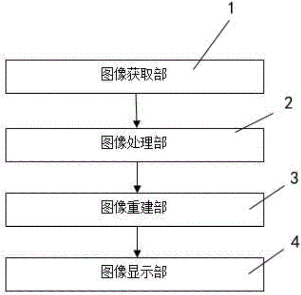
[0039] Please refer to figure 1 , figure 1 It is a structural block diagram of a device for examining rectal-anal mucosal lesion tissue of the present invention. A device for examining rectal-anal mucosal lesion tissue, the device includes an image acquisition unit 1, an image processing unit 2, an image reconstruction unit 3, and an image display unit 4; the image acquisition unit 1 is used to acquire rectal - the image of the anal canal mucosa; the image processing unit 2 is used to process the image data of the image acquisition unit 1, and the image reconstruction unit 3 is used to regenerate the rectum-anal canal captured by the image acquisition unit 1 Mucous membrane image; the image display unit 4 is used to display the image generated by the image reconstruction unit 3 .

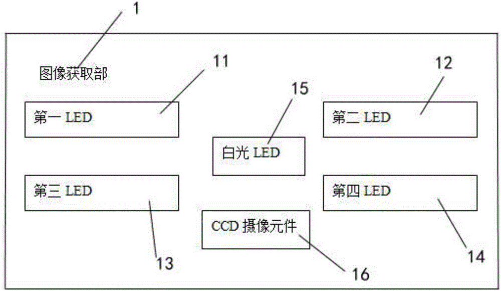
[0040] Please refer to figure 2 , figure 2It is a schematic diagram of the ...

Embodiment 2

[0045] Example 2 The device of the present invention detects the glandular duct typing opening of the lesion

[0046] The duct opening of the lesion adopts Kudo Shinhide Nagata's classification, and there are 5 types in total, as follows:

[0047] Type I: It is round and is the duct opening of normal mucosa, but it is of great significance for the diagnosis of submucosal tumors. Submucosal tumors originate from the submucosa or muscular layer of the tumor, and the type of duct opening usually does not change, so Often this type is gland duct opening.

[0048] Type II: star-shaped or papillary, the opening is larger than that of normal glandular ducts, and its tissue shows proliferative lesions. This type of duct opening can be diagnosed as a hyperplastic lesion once it is found under a magnifying glass. According to relevant reports, the diagnostic accuracy rate is as high as 100%.

[0049] III L Type: The opening of the gland duct is tubular or almost round, larger than th...

Embodiment 3

[0060] Example 3 The device of the present invention detects flat and depressed lesions

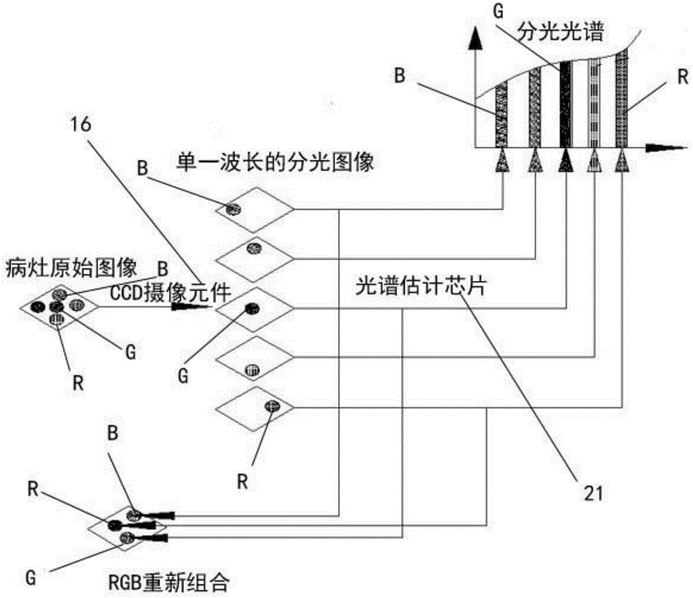
[0061] After the reflected light image of the lesion illuminated by the first LED 11 is processed by the CCD imaging element 16, the wavelength distribution is: R560nm, G440nm, B420nm; the reflected light image of the lesion illuminated by the second LED 12 is processed by the CCD imaging element 16. The wavelength distribution is: R555nm , G445nm, and B415nm were used to observe the lesions, and it was found that the boundary between the flat and depressed lesions and the surrounding mucosa was clear, the outline of the lesions was prominent, the tumor blood vessels dilated at the edge of the lesions, the surface of the lesions was uneven, the color of the depressions became lighter, and the vascular network was destroyed. At the same time, conventional endoscopy was used to observe, and it was found that the boundary between flat and depressed lesions and the surrounding mucosa was not c...


PUM


Abstract

The invention relates to a device for examining rectum-anal canal mucosal lesion tissues. The device comprises an image acquisition portion, an image processing portion, an image reconstruction portion and an image display portion. The image acquisition portion is used for acquiring rectum-anal canal mucosal images; the image processing portion is used for processing image data acquired by the image acquisition portion; the image reconstruction portion is used for reconstruction of the rectum-anal canal mucosal images shot by the image acquisition portion; the image display portion is used for displaying the images generated by the image reconstruction portion. The device has advantages that the contrast ratio of surface blood capillary and subcutaneous microvascular imaging is increased, the color tone of lesions is deeper, boundaries of the lesions and surrounding normal mucosa are clearer, blood vessel morphological changes and texture images can be observed clearly, and accordingly direct references can be provided for doctors to recognize the lesions, judge benign or malignant tumors and the like, diagnosis sensitivity can be improved, accuracy in lesion range judgment is achieved, and corresponding surgical operations can be guided.

Description

technical field [0001] The invention relates to the technical field of biomedical engineering, in particular to a device for examining rectum-anal canal mucosal lesion tissue. Background technique [0002] The anorectal canal area has an important function of controlling fecal excretion. Due to its complex structure and various tissue types, it is prone to morphological abnormalities and functional diseases, such as hemorrhoids, abscesses, and rectal cancer. With the continuous improvement of people's living standards and changes in eating habits, rectal cancer is growing rapidly. Rectal cancer is a common malignant tumor of the digestive tract in my country. It is estimated to increase at a rate of 4% every year, and the incidence rate ranks first among malignant tumors. four. According to statistics, the incidence rate of rectal cancer in Shanghai in 1999 was 18.3 / 100,000 males and 13.9 / 100,000 females. [0003] In the prior art, methods commonly used for rectal-anal muco...

Claims


Application Information

Patent Timeline
IPC IPC(8): A61B1/31A61B1/04A61B1/06A61B1/00A61B5/00
CPCA61B1/31A61B1/00009A61B1/00131A61B1/04A61B1/0684A61B5/004A61B5/0075A61B5/0084
Inventor 钱欢徐宏智
Owner THE FIFTH PEOPLES HOSPITAL OF SHANGHAI
Why Eureka
Try Eureka