Method for visually marking genome sites

A genome and labeling technology, applied in the biological field, can solve problems such as the inability to accurately label genomic elements and special sites in human cells, low signal-to-noise ratio, and genome damage.

Active Publication Date: 2017-06-06
INSITUTE OF BIOPHYSICS CHINESE ACADEMY OF SCIENCES
View PDF2 Cites 2 Cited by
  • Summary
  • Abstract
  • Description
  • Claims
  • Application Information

AI Technical Summary

Problems solved by technology

Although a variety of genome visualization labeling technologies have been used in scientific research, the fluorescent labeling Lac or Tet system needs to integrate a large exogenous fragment of about 10kb into the target site of the genome, so there are problems such as low labeling efficiency and potential genome damage ( Robinett,C.C.,etal.,In vivo localization of DNA sequences andvisualization of large-scale chromatin organization using lac operator / repressor recognition.J Cell Biol,1996.135(6Pt 2):p.1685-700.2.Heun,P.,etal. ,Chromosome dynamics in the yeast interphase nucleus.Science,2001.294(5549):p.2181-6.); Fluorescence in situ hybridization (FISH) is currently the gold standard for studying the location of specific sequences in the genome, but this method requires chemical The operation is performed in fixed cells, so genome visualization in living cells cannot be achieved (Levsky, J.M. and R.H. Singer, Fluorescence in situ hybridization: past, present and future. J Cell Sci, 2003.116 (Pt 14): p.2833-8 .); Although the CRISPR / Cas9 technology that has emerged in recent years can achieve precise labeling of special genomic sites such as telomeres in living cells, the high background in the nucleus leads to low signal-to-noise ratios and complex systems. Accurate labeling of genomic loci in other human cells (such as pluripotent stem cells and terminally differentiated cells) other than cell lines (Levsky, J.M. and R.H. Singer, Fluorescence in situ hybridization: past, present and future. J Cell Sci , 2003.116(Pt 14): p.2833-8.); Another transcription activator-like effector element (TALE) used as gene editing technology can also be used for visual labeling of genomic loci, however existing Reports neither validated the correctness of TALE-mediated genomic labeling with the FISH gold standard, but also showed strong heterogeneity of labeling in different cell types (Ma, H., P. Reyes-Gutierrez, and T. Pederson, Visualization of repetitive DNA sequences in human chromosomes with transcription activator-like effectors.Proc NatlAcad Sci USA, 2013.110(52):p.21048-53.)
[0004] In summary, the existing genome visualization labeling technologies are flawed and cannot meet the needs of accurately labeling human cell genome elements and special sites

Method used


Image

  • Method for visually marking genome sites
  • Method for visually marking genome sites
  • Method for visually marking genome sites

Examples


Embodiment 1

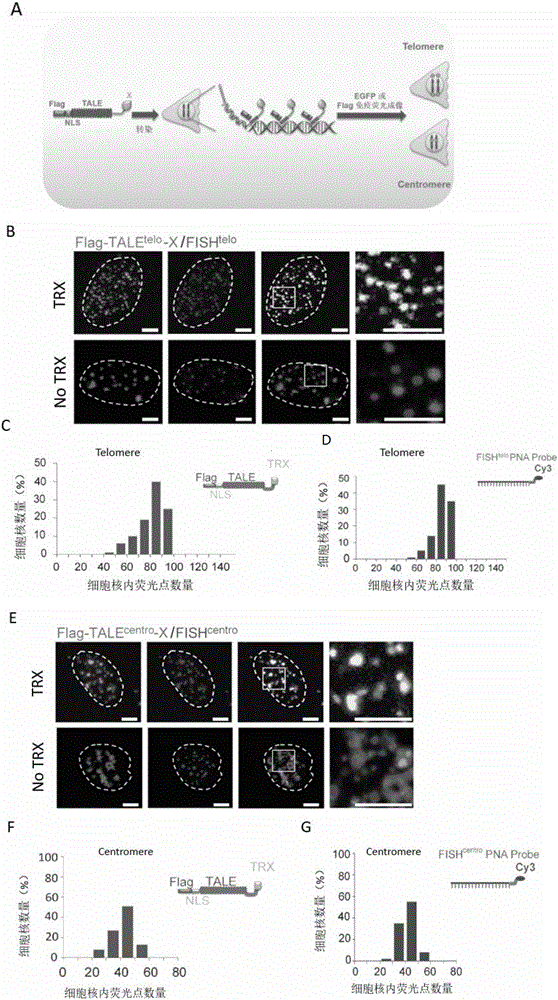
[0098] Example 1, Establishment of Genome Visualization Labeling Technology (TTALE) for Accurately Marking Telomere and Centromere in Cell Genome

[0099] 1. Construction of the expression vector TTALE for fusion expression of TALE and TRX

[0100] 1. TALE vectors that recognize telomeres and centromeres

[0101] Refer to the literature "Zhang, F., et al., Efficient construction of sequence-specific TAL effector for modulating mammalian transcription. Nat Biotechnology, 2011.29(2): p.149-53, using the method in Golden Gate Assembly" using TALE Toolbox Kit (Addgene, USA, Cat. No. 1000000019) to construct TALE vectors recognizing telomeres and TALE vectors recognizing centromeres, respectively.

[0102] (1) TALE carrier that recognizes telomeres

[0103] The nucleotide sequence of the TALE vector recognizing telomeres is shown in Sequence 1. Telomere-recognizing TALE vector expresses fusion protein TALE telo , the fusion protein TALE tel The target sequence of the identifie...

Embodiment 2

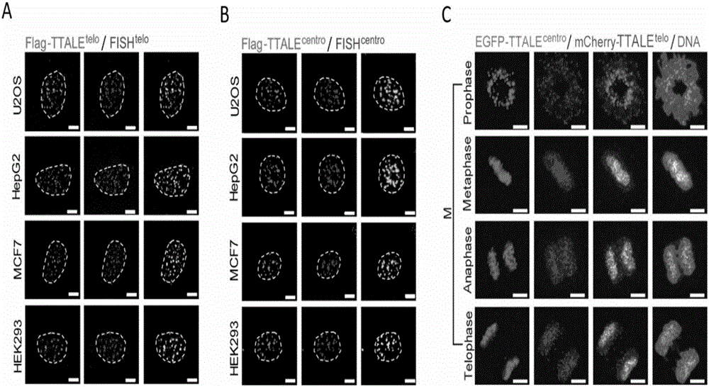
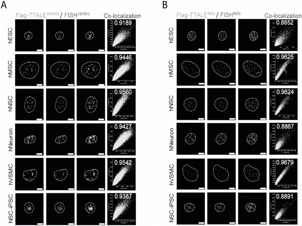
[0119] Example 2, Application of TTALE in Accurately Marking Telomere, Centromere, Nucleolus Organization Region Ribosomal RNA Coding Sequence (NOR-rDNA) and Coding Gene Locus (MUC4) in Different Types of Human Cells

[0120] 1. Application of TTALE in precise labeling of telomeres and centromeres in different types of human cells

[0121] The U2OS cells in Example 1 were replaced with the following cells respectively: human tumor cell lines (MCF7 and HepG2, American ATCC Company, product numbers HTB-22 and HB-8065 respectively), human embryonic kidney cell lines (HEK293, American ATCC Company , product number is CRL-1573), human pluripotent stem cells (embryonic stem cells hESC, Wicell Company, product number WA-09), human pluripotent stem cells (induced pluripotent stem cells iPSC, Wicell Company, product number IISH6i-CML17), adult stem cells (Mesenchymal stem cells, Lonza company, product number is PT-2501), adult stem cells (neural stem cells hNSC, Wicell company, product...

Embodiment 3

[0151] Example 3. Application of TTALE to mark telomeres in different cell aging models in visualizing the dynamic changes of cell aging process

[0152] 1. Preparation of TTALE vector for telomere recognition of EGFP fusion expression

[0153] The EGFP coding gene sequence shown in the 7382-8098 position of sequence 13 is replaced with the TTALE carrier (mCherry-TTALE telo ) in the mCherry coding gene sequence to obtain the TTALE carrier (EGFP-TTALE telo ), the N-terminus of the fusion protein has a FLAG tag sequence and a nuclear localization sequence NLS.

[0154] 2. Human mesenchymal stem cells of different aging models labeled with TTALE carrier expressing EGFP fusion and recognizing telomeres

[0155] (1) Establishment of human mesenchymal stem cells in different aging models

[0156] 1) Preparation of wild-type human mesenchymal stem cells (WT-MSC) and WRN gene-deleted human mesenchymal stem cells (WS-MSC)

[0157] In the present invention, wild-type human embryonic...


PUM


Abstract

The invention discloses a method for visually marking genome sites. According to the method, a novel visual genome marking tool TTALE is established by fusing thioredoxin (TRX) at the c-terminal of the traditional transcriptional activation-like effect element (TALE) protein. Experiments prove that the TTALE can be used for accurately marking genome repetitive sequences such as telomere, centromere and ribosomal ribonucleic acid (RNA) coded sequences (ribosomal deoxyribonucleic acid (rDNA)) in different types of human cells such as a tumor cell line, embryonic stem cells, adult stem cells and terminally differentiated cells, and coded genetic loci (MUC4). A TTALE technology can make up for the lack of accurate, simple, convenient and long-acting live cell genome visualization marking techniques in the current scientific research and clinical markets, thereby promoting the development of basic research and clinical diagnosis and treatment of human senility and vital diseases.

Description

technical field [0001] The invention belongs to the field of biotechnology, and in particular relates to a method for visually marking genome sites. Background technique [0002] In the nucleus of human cells, about 3.2 billion base pairs form the huge human genome, which is further condensed to form 23 pairs of chromosomes of different sizes. Although the completion of the Human Genome Project has helped us obtain all the sequence information of the human genome, this is only for us to understand the structure of the human genome, and understand the three-dimensional structure of chromatin composed of the genome and important biological processes such as human development, aging and disease The first step in a relationship. In fact, many studies have proved that genome structure disorder is an important factor leading to aging and several serious diseases (Zhang, W., et al., Aging stem cells. A Werner syndrome stem cell model unveils heterochromatinalterations as a driver ...

Claims


Application Information

Patent Timeline
Patent Type & Authority Applications(China)
IPC IPC(8): C07K19/00C12N15/62C12N15/85C12N15/65
CPCC07K14/195C07K2319/09C07K2319/35C07K2319/60C12N15/65C12N15/85C12N2800/107
Inventor 刘光慧曲静任若通邓丽萍
Owner INSITUTE OF BIOPHYSICS CHINESE ACADEMY OF SCIENCES
Why Eureka
Try Eureka