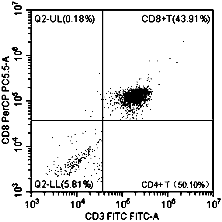
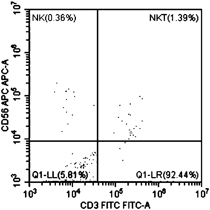
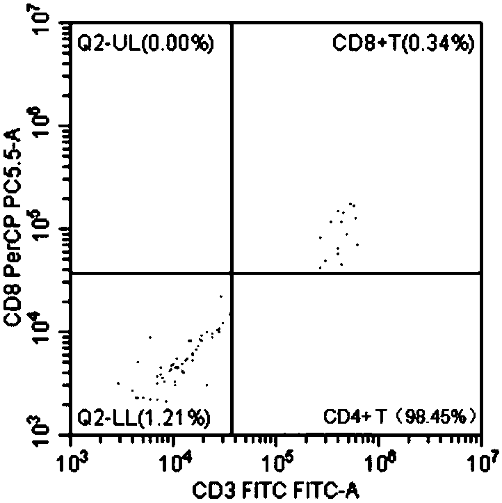
Identification method and application of eb virus-infected lymphocyte subpopulation
A lymphocyte and EB virus technology, applied in the field of medical detection, can solve the problems of poor EB virus detection and other problems
- Summary
- Abstract
- Description
- Claims
- Application Information
AI Technical Summary
Problems solved by technology
Method used
Image
Examples
Embodiment 1
[0045] This embodiment provides a magnetic bead-based identification method for EB virus-infected lymphocyte subsets, which includes the following steps:
[0046] 1. Extract human peripheral blood mononuclear cells (HPBMC): 6mL fasting venous blood, placed in a sterile test tube with EDTA anticoagulation, diluted 1 times with PBS, and separated PBMC with lymphocyte separator (2000 turns 20 minutes), using Centrifuge time with buffer solution (MACS BSA Stock Solution (#130-091-376) and autoMACS Rinsing Solution (#130-091-222) 1:20 ratio), and place in a refrigerator at 4 degrees;
[0047] 2. Aspirate the middle tunica albuginea layer, add 10mL PBS, 1400rpm, 10min to wash and precipitate PBMC;
[0048] 3. Discard the supernatant, add 2mL red blood cell lysate (Solebold red blood cell lysate item number: Cat#R1010) for lysis, mix well, and incubate at room temperature for 5 minutes;
[0049] 4. After mixing well, draw 20μL of cells and dilute 1:3 with 2% glacial acetic acid, then take th...
Embodiment 2
[0080] This embodiment provides a flow cytometer-based identification method for EB virus-infected lymphocyte subsets, which includes the following steps:
[0081] 1. Take 6 mL of peripheral blood, anticoagulate with heparin, dilute to 12 mL with PBS, and mix;
[0082] 2. Slowly add the diluted blood along the test tube wall to the surface of the 15mL lymphocyte separation solution. Do not use too much force to avoid mixing the blood and the separation solution and maintain a clear layered state;
[0083] 3. Centrifuge at 2000 rpm for 20 minutes at 18-20°C. After centrifugation, the blood in the test tube can be clearly divided into 4 layers. The upper layer is the plasma layer, and the middle layer is the separation layer (mononuclear cells are located between the plasma layer and the separation layer) , The bottom layer is the red blood cell layer, and the red blood cell layer is the granulocyte layer;
[0084] 4. Aspirate the lymphocyte layer between the upper layer and the middle ...
PUM
 Abstract
Description
Claims
Application Information
 - R&D
- Intellectual Property
- Life Sciences
- Materials
- Tech Scout
- Unparalleled Data Quality
- Higher Quality Content
- 60% Fewer Hallucinations
Browse by: Latest US Patents, China's latest patents, Technical Efficacy Thesaurus, Application Domain, Technology Topic, Popular Technical Reports.
© 2025 PatSnap. All rights reserved.Legal|Privacy policy|Modern Slavery Act Transparency Statement|Sitemap|About US| Contact US: help@patsnap.com


