Method for quantitatively analyzing cell DNA damage caused by nitrosamine or nitrosamine metabolite through high-content technology

A quantitative analysis and nitrosamine technology, applied in the direction of material analysis, material excitation analysis, material analysis through optical means, etc., can solve the problems of cumbersome operation, large error, and inability to provide intracellular fluorescence focus distribution, etc., to achieve cell The effect of small volume and small sample volume

Inactive Publication Date: 2017-07-25
CHINA NAT TOBACCO QUALITY SUPERVISION & TEST CENT +1
View PDF5 Cites 1 Cited by
  • Summary
  • Abstract
  • Description
  • Claims
  • Application Information

AI Technical Summary

Problems solved by technology

Flow cytometry and Western Blot are cumbersome to operate, and adherent cells need to be enzymatically hydrolyzed into single-cell suspension before detection, which destroys the structural and functional integrity of cells, and the detection throughpu

Method used


Image

  • Method for quantitatively analyzing cell DNA damage caused by nitrosamine or nitrosamine metabolite through high-content technology
  • Method for quantitatively analyzing cell DNA damage caused by nitrosamine or nitrosamine metabolite through high-content technology
  • Method for quantitatively analyzing cell DNA damage caused by nitrosamine or nitrosamine metabolite through high-content technology

Examples


Embodiment 1

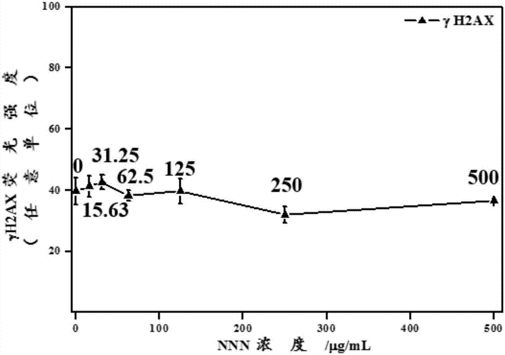
[0065] The γH2AX induced by NNN was measured after 24h exposure.

[0066] A549 cells in logarithmic growth phase were collected, planted in 96-well plates at 10,000 cells per well, in RPMI-1640 medium containing 10% FBS and 2mmoL / L L-glutamine at 37°C, 5% CO 2 Incubate in the incubator for 24h.

[0067] The cell culture fluid after culturing for 24 h was discarded, and the cell culture fluid containing 0, 15.63, 31.25, 62.5, 125, 250 and 500 μg / mL NNN (also containing 10% FBS and 2mmoL / L L-glutamine RPMI-1640 culture medium) was used as the cell poisoning solution, and the cells were continued to be cultured for 24 h.

[0068] After the poisoning, discard the cell poisoning solution, add 100 μL PBS to each well and wash twice for at least 5 minutes each time; then add 50 μL 4% paraformaldehyde solution to each well and fix at room temperature for 15 minutes; fix the cells with PBS buffer Wash twice for at least 5 min each time; then add 50 μL of 0.5% Triton-100X solution (in...

Embodiment 2

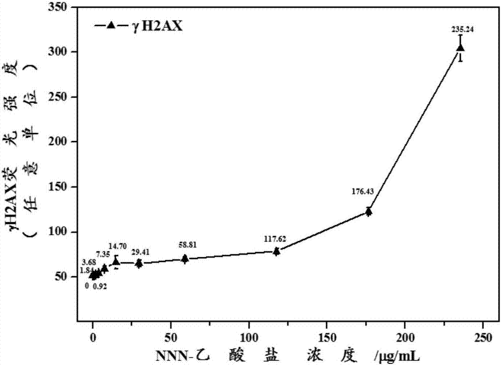
[0073] γH2AX induced by NNN-acetate exposure for 24 h was measured.

[0074] The experimental procedure was carried out as described in Example 1, the only difference was that cells were cultured with 0, 0.92, 1.84, 3.68, 7.35, 14.70, 29.41, 58.81, 117.62, 176.43 and 235.24 μg / mL NNN-acetate liquid as a cell venom.

[0075] image 3Shown is the dose-effect relationship curve of γH2AX produced by A549 cells induced by different concentrations of NNN-acetate 24 hours after exposure. It can be seen from the figure that with the increase of the concentration of NNN-acetate, the γH2AX produced in the cells induced by it shows a significant dose-dependent increase, which has a significant dose-effect relationship. When the concentration of NNN-acetate was 117.62 μg / mL, the induced γH2AX was 1.5 times higher than that of the normal group (that is, when the concentration of NNN-acetate was 0).

Embodiment 3

[0077] The γH2AX induced by NNK after 24h exposure was measured.

[0078] The experimental procedure was carried out as described in Example 1, with the only difference that cell culture solutions containing 0, 15.63, 31.25, 62.5, 125, 250 and 500 μg / mL NNK were used as cell poisoning solutions.

[0079] Figure 4 Shown is the dose-effect relationship curve of γH2AX produced by A549 cells induced by different concentrations of NNK 24 hours after exposure. It can be seen from the figure that with the increase of NNK concentration, the γH2AX produced by A549 cells has no significant change compared with the normal group (that is, when the NNK concentration is 0).


PUM


Abstract

The invention discloses a method for quantitatively analyzing cell DNA damage caused by nitrosamine or nitrosamine metabolite through a high-content technology. The method comprises 1) cell culture, 2) cell exposure to toxicant, 3) gamma H2AX immunofluorescence labeling, and 4) quantitative analysis based on a high-content technology. The method has the advantages that the high-content imaging system is used for automatic imaging and the DNA double-strand breaking marker gamma H2AX protein produced by induction of nitrosamine or nitrosamine metabolite is quantitatively analyzed so that the direct and rapid detection of cells are realized and sample treatment is convenient, the high-resolution imaging realizes direct observation of distribution of gamma H2AX in a cell nucleus, the image data can be easily stored and re-analyzed, the method can realize quantitative analysis of gamma H2AX induced by nitrosamine or nitrosamine metabolite in each cell nucleus, and the detection result is sensitive and accurate.

Description

technical field [0001] The invention belongs to the technical field of in vitro detection of DNA damage, and more specifically relates to an in vitro quantitative analysis method for cellular DNA damage caused by nitrosamines or nitrosamine metabolites. Background technique [0002] Tobacco-specific nitrosamines, such as NNN and NNK, are unique and typical N-nitrosamines in tobacco and smoke. There is sufficient evidence to show that NNK and NNN are strong rodent carcinogens. The electrophilic intermediates produced by NNN and NNK in the metabolic process can form adducts with DNA and remain in the nasal mucosa, lung, liver, and pancreas tissues of rodents, thereby inducing canceration of related target tissues. At present, the International Organization for Research on Cancer (IARC) under the World Health Organization (WHO) has listed tobacco-specific nitrosamines NNN and NNK as Class 1 carcinogens, and the World Health Organization Framework Convention on Tobacco Control (...

Claims


Application Information

Patent Timeline
IPC IPC(8): G01N21/64
CPCG01N21/6486
Inventor 侯宏卫张森胡清源陈欢王安刘勇
Owner CHINA NAT TOBACCO QUALITY SUPERVISION & TEST CENT
Why Eureka
Try Eureka