Schistosoma japonicus detection method and kit and primer thereof

A detection method and technology for schistosomiasis, applied in the field of detection of parasitic disease transmission vectors, can solve the problems of inability to manufacture, long time, unsuitable loose stools, etc., and achieve the effects of an effective diagnostic approach, strong specificity and high sensitivity

Active Publication Date: 2017-08-18
NANJING MUNICIPAL CENT FOR DISEASE CONTROL & PREVENTION
View PDF5 Cites 2 Cited by
  • Summary
  • Abstract
  • Description
  • Claims
  • Application Information

AI Technical Summary

Problems solved by technology

[0004] However, there are still many imperfections in the existing detection methods. For example, the most common direct smear method is to detect through morphological identification. In actual operation, the components in the feces are different. It is difficult to distinguish and identify fungal spores with similar shapes, different insect eggs and other microorganisms, that is to say, whether the results of this method are accurate or not is highly dependent on the experience of the inspectors in morphological identification; the advantages of microscopic examination technology are simple technology, easy operation, The disadvantage is that it is not suitable for loose stools, and the detection rate is reduced, especially the improved Kato thick smear method, which cannot produce satisfactory films for loose stools
In addition, in order to improve the detection rate, in actual operation, it is often necessary to perform operations such as elutriation, concentration, and miracidium hatching on feces, which takes a long time, and this operation has high biosafety requirements, and grassroots units often do not meet biosafety requirements. The operation will pose a direct threat to the health of the operator

Method used


Image

  • Schistosoma japonicus detection method and kit and primer thereof
  • Schistosoma japonicus detection method and kit and primer thereof
  • Schistosoma japonicus detection method and kit and primer thereof

Examples


Embodiment 1

[0049] Extraction method of Schistosoma japonicum DNA

[0050] 1. Preparation of feces samples: Take a small amount of freshly collected feces, about 0.5 grams, put it in a 1.5 ml centrifuge tube, add 1 ml of normal saline, shake fully on the shaker, centrifuge at 14000 g for 30 seconds, discard the supernatant; store in alcohol or feces in other solutions, repeat washing 3 times, and discard the supernatant.

[0051] 2. Lysis: Add 200 microliters of lysate solution (4M urea, 200mM Tris, 20 mM NaCl, 200mM EDTA, pH 7.4) and 40 microliters of proteinase K (1mg / mL) to the precipitate, mix quickly, and incubate at 55 degrees for 0.5 -1 hour until the liquid is clear. Blow back and forth 3 times with a 1ml syringe (needle removed).

[0052] 3. Nucleic acid precipitation: add 200 microliters of binding solution (6M guanidine hydrochloride, 10M urea, 10mM Tris-HCL, 20% TritonX-100 (v / v), pH 4.4), mix immediately, and incubate at 70 degrees for 10 minutes. Add 100 µl of isopropanol...

Embodiment 2

[0059] Loop-mediated isothermal DNA amplification: Take three 0.2mL PCR thin-walled tubes and mark them as positive control tubes, detection tubes, and negative control tubes;

[0060] Add the following ingredients to each tube in turn:

[0061] Reaction mixture: 15mM Tris–HCl (pH8.6), 1.6mM dNTPs, 8mM KCl, 25mM Calcein, 0.5mM MnCl 2 , 12mM (NH 4 ) 2 SO 4 , 10mM MgSO 4 , 1.0M Betaine (Sigma–Aldrich) and Tween 20 at 0.13% by volume;

[0062] 16U of Bst DNA polymerase (New England Biolabs);

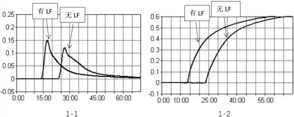
[0063] Mixed primers: 0.2 μM F3 and 0.2 μM B3, 1.4 μM FIP, 1.4 μM BIP and 1.0 μM LF; the nucleotide sequences of F3, B3, FIP, BIP and LF are shown in SEQ ID NO: 1-5 .

[0064] Add 5 μL of the DNA prepared in Example 1 to the detection tube; add 5 μL of deionized water negative control to the negative control tube; add the positive control of standard schistosome DNA to the positive control tube.

[0065] After a brief centrifugation, place it in a constant temperature water bath at ...

Embodiment 3

[0067] Loop-mediated isothermal DNA amplification: Take three 0.2mL PCR thin-walled tubes and mark them as positive control tubes, detection tubes, and negative control tubes;

[0068] Add the following ingredients to each tube:

[0069] Reaction mixture: 25mM Tris–HCl (pH9.0), 1.2mM dNTPs, 8mM KCl, 100μM hydroxynaphthol blue, 8mM (NH 4 ) 2 SO 4 , 6mM MgSO 4 , 0.6M betaine (Sigma–Aldrich) and Tween20 with a volume percentage of 0.07%;

[0070] 16U of Bst DNA polymerase (New England Biolabs);

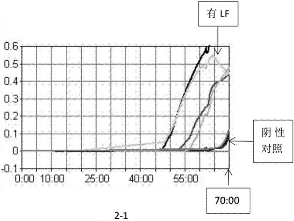
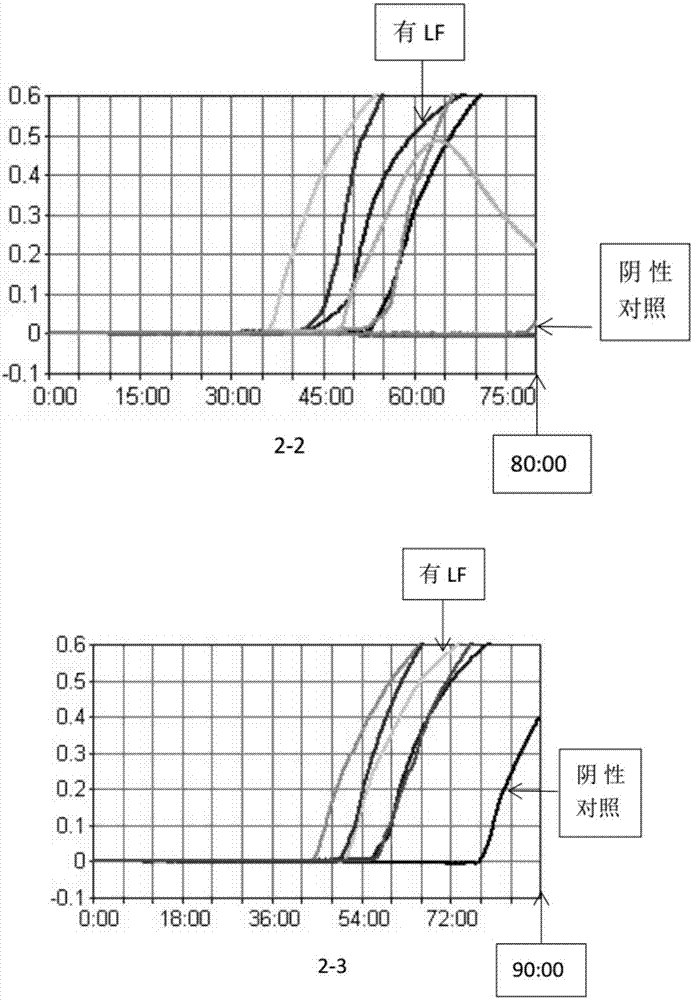
[0071] Mixed primers: 0.2 μM F3 and 0.2 μM B3, 1.8 μM FIP, 1.8 μM BIP and 0.6 μM LF; the nucleotide sequences of F3, B3, FIP, BIP and LF are shown in SEQ ID NO: 1-5 .

[0072] Add 5 μL of the DNA prepared in Example 1 to the detection tube; add 5 μL of deionized water negative control to the negative control tube; add the positive control of standard schistosome DNA to the positive control tube.

[0073] After a brief centrifugation, place it in a constant temperature water bath a...


PUM


Abstract

The invention relates to the field of parasitic disease media detection, in particular to a schistosoma japonicus detection method and a kit and a primer thereof. The kit comprises an LAMP primer set, a reaction mixture and a DNA polymerase. The reaction mixture contains hydroxynaphthol blue as an indicator. The LAMP primer set comprises an outer primer pair formed by F3 and B3 and an inner primer pair formed by FIP and BIP; the LAMP primer set further comprises an LF loop primer. The kit utilizing the detection method can detect the schistosoma japonicas efficiently, quickly and specifically.

Description

technical field [0001] The invention relates to the field of detection of parasitic disease transmission media, in particular to a method for detecting schistosomiasis eggs and a kit and primers used in the method. Background technique [0002] Schistosoma is also called schistosoma. Schistosoma parasitizes most vertebrates, and the eggs pass through the vein wall into the bladder and are excreted with urine. The larvae develop inside the intermediate host snail. Mature larvae enter the final host through the skin or mouth. Schistosoma mansoni (S. mansoni, namely Schistosoma mansoni) is mainly distributed in Africa and northern South America in the large and small intestinal veins. Eggs are excreted in feces. The larva enters the snail and returns to the final host through the skin. Schistosoma japonicum (S. japonicum, namely Schistosoma japonicum) is mainly found in mainland China, Japan, Taiwan, the East Indies and the Philippines. In addition to humans, it also attac...

Claims


Application Information

Patent Timeline
Patent Type & Authority Applications(China)
IPC IPC(8): C12Q1/68C12Q1/04C12N15/11C12R1/90
CPCC12Q1/686C12Q1/6893C12Q2531/113C12Q2521/101
Inventor 张洪英杨佩才谢国祥
Owner NANJING MUNICIPAL CENT FOR DISEASE CONTROL & PREVENTION
Why Eureka
Try Eureka