Method for detecting single biological marker by using total internal reflection fluorescent microscope

A biomarker and fluorescence microscope technology, applied in the field of biological detection, can solve the problems of unsure of the treatment method, missing the best treatment opportunity, and exacerbation of the disease in sick animals, and achieves convenient and easy detection steps, short detection time, and low background low noise effect

Inactive Publication Date: 2017-08-29
NANJING AGRICULTURAL UNIVERSITY
View PDF4 Cites 2 Cited by
  • Summary
  • Abstract
  • Description
  • Claims
  • Application Information

AI Technical Summary

Problems solved by technology

There are many methods for detecting pathogens, such as enzyme-linked immunosorbent assay (ELISA), Western blot (Western Blot) and polymerase chain reaction (PCR), etc., but these methods have more or less problems, such as operation Disadvantages such as complexity, high cost, and low sensitivity
It is difficult to detect trace pathogens in biological tissues or cells using conventional methods, which makes it impossible to determine the best treatment for sick animals, or even miss the best time for treatment, leading to aggravation of the disease

Method used


Image

  • Method for detecting single biological marker by using total internal reflection fluorescent microscope
  • Method for detecting single biological marker by using total internal reflection fluorescent microscope
  • Method for detecting single biological marker by using total internal reflection fluorescent microscope

Examples


Embodiment 1

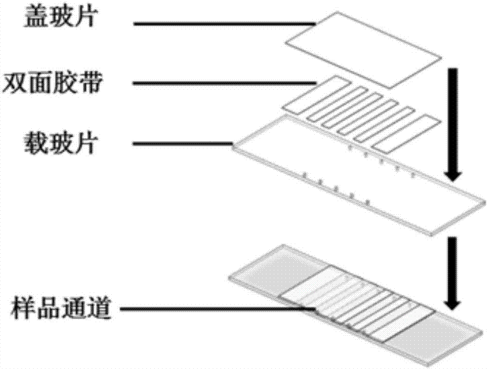
[0048] Embodiment 1 is made to be used for the sample small dish of sample addition

[0049] 1. Use an ordinary drill to drill 10 holes with a diameter of 0.8mm on a glass slide with a thickness of 1mm. This hole is very suitable for adding samples with a 200ul yellow tip;

[0050] 2. Rinse the slides repeatedly with deionized water;

[0051] 3. Ultrasonic cleaning of slides and coverslips with an acetone / alcohol solution with a volume ratio of 1:1 for 20 minutes;

[0052] 4. Rinse three times with deionized water;

[0053] 5. Ultrasonic cleaning with 1mol / L potassium hydroxide solution for 20 minutes;

[0054] 6. Rinse three times with deionized water;

[0055] 7. Dry the slides and coverslips in an oven;

[0056] 8. Place the slide and cover slip in a mixture containing 250ml of methanol, 10ml of anhydrous acetic acid, and 2ml of aminosilane coupling agent, sonicate for 1min, then let it stand for 20min to react;

[0057] 9. Place the slide and cover slip in methanol fo...

Embodiment 2

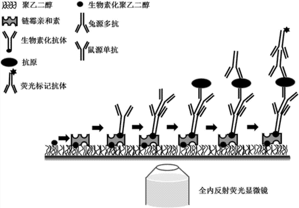
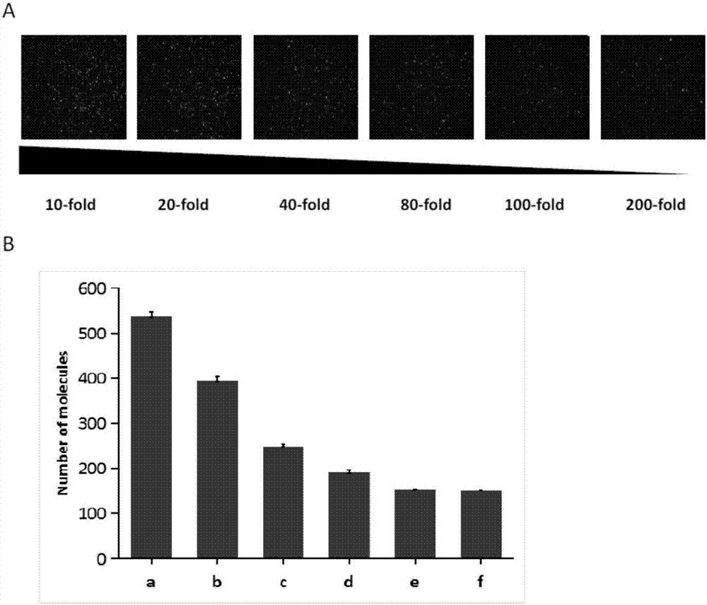
[0065] Example 2 Step-by-step loading of antibodies and antigens to be detected Immobilize pathogenic molecules, and use a fluorescent inverted microscope based on total internal reflection to obtain the number of pathogenic molecules

[0066] 1. Take 10ul of 0.2mg / mL streptavidin dissolved in T50 (10mmol / L Tris-HCl, (pH 8.0) and 50mmol / L NaCl) solution and add it to the sampling channel of the sample dish, and incubate at room temperature for 5min; There will be a connection between biotin and avidin;

[0067] 2. Take 20ul of T50 solution and add it to the sampling channel to wash away unbound streptavidin;

[0068] 3. Add 10ul of 40nM biotin-labeled goat anti-mouse secondary antibody to the loading channel, and incubate at room temperature for 15 minutes; the connection between biotin and avidin will occur, and the secondary antibody will be immobilized on the surface of the channel;

[0069] 4. Add 20ul of T50-BSA (0.1mg / ml bovine serum albumin (BSA) dissolved in T50 solut...


PUM

PropertyMeasurementUnit
refractive indexaaaaaaaaaa

Abstract

The invention discloses a method for detecting a biological marker by using a total internal reflection fluorescent microscope. The method comprises the following steps of utilizing biotin-avidin connection, and antigen and antibody reaction or nucleic acid molecule and probe reaction, using a total internal reflection fluorescent exciting type to excite a detection molecule of a fluorescent marker which is fixed in the 100nm range of the carrier surface, and collecting a fluorescent molecule signal by the fluorescent microscope, so as to obtain the number of to-be-detected biology markers. The method has the advantages that because the free fluorescent molecules in the solution are not excited, the background noise of the collected fluorescent signal is lower, and the signal to noise ratio is very high; the fluorescent signal of the single molecule can be distinguished, so that the detection sensitivity is high; the detection method can be used for detecting various types of biological markers, and an important meaning is realized for the finding of pathogen in diseases and the preventing and controlling of diseases.

Description

technical field [0001] The invention relates to the technical field of biological detection, in particular to a method for detecting a single biological marker using an inverted microscope based on total internal reflection fluorescence. Background technique [0002] Many diseases caused by pathogenic microorganisms occur and spread widely in the world today, seriously endangering the health of humans, animals and plants, and affecting the quality of human life. Understanding the properties of pathogenic microorganisms is essential for disease prevention and treatment. There are many methods for detecting pathogens, such as enzyme-linked immunosorbent assay (ELISA), Western blot (Western Blot) and polymerase chain reaction (PCR), etc., but these methods have more or less problems, such as operation Complexity, high cost, low sensitivity and other disadvantages. It is difficult to detect trace pathogens in biological tissues or cells using conventional methods, which makes ...

Claims


Application Information

Patent Timeline
Patent Type & Authority Applications(China)
IPC IPC(8): G01N21/64
CPCG01N21/6428
Inventor 刘斐吴诚诚周继勇王国亚邢刚雷静
Owner NANJING AGRICULTURAL UNIVERSITY
Why Eureka
Try Eureka