

Application of near-infrared luminescent ruthenium complex to cell pH sensing
A dual-nuclear ruthenium complex, cell technology, applied in the field of cell pH sensing, can solve problems such as rare application reports
- Summary
- Abstract
- Description
- Claims
- Application Information
AI Technical Summary
Problems solved by technology
Method used
Image

Examples
Embodiment 1
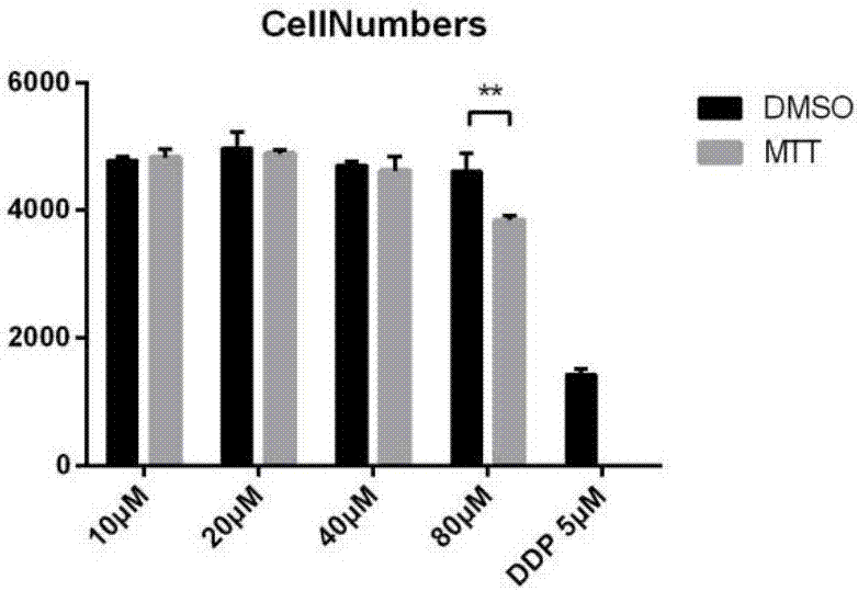
[0016] Embodiment 1: cell culture and cytotoxicity experiment
[0017] Cervical cancer HeLa cells were cultured with cell culture medium at 370°C in CO 2 cultured in an incubator (95% relative humidity, 5% CO 2 ). The in vitro cytotoxicity of the complex was determined on a high-content analysis system produced by Molecular Devices. Take cervical cancer HeLa cells that are in the exponential growth phase, make cell suspension with culture medium, and count with a cell counting plate, according to 6×10 3 Cells / well were seeded in 96-well plates for subculture. After 24 hours of cell subculture, add the dimethyl sulfoxide solution of the complex to make the final concentration 10-80 μM. 100 μL of medium and cisplatin were used as negative and positive controls, respectively. The 96-well plate was placed in an incubator with 95% relative humidity and 5% carbon dioxide at 37°C for 48 hours. Then stained with 10 μg / mL Hoechst 33342 and 1 μg / mL propidium iodide PI in phosphate b...
Embodiment 2
[0019] Example 2: Flow Cytometry Analysis
[0020] Cervical cancer HeLa cells were digested with trypsin, suspended in 5 mL of phosphate buffer, and centrifuged at 1000 rpm for 10 minutes. Discard the supernatant and add 1 mL of phosphate buffer to resuspend the cells. A sample containing 20000 cells was analyzed by a NovoCyte type flow cytometer (ACEA, USA). The excitation wavelength is 488nm, and the emitted photons are collected through a 780 / 20nm filter.
[0021] We explored the optimal concentration of the complexes and the optimal incubation time by flow cytometry. It can be seen from Figure 3 that when the concentration of the complex is 80 μM, the luminous intensity of the cells is still not saturated. Due to the limitation of the solubility of the complex, we choose a concentration of 40 μM complex close to the maximum luminous intensity of the cells in our experiment. It can be seen from Figure 4 that when the incubation time reaches 8 hours, the cells reach the m...
Embodiment 3
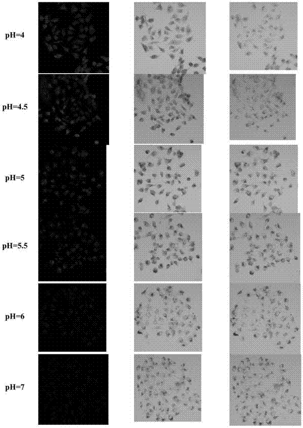
[0022]Example 3: HeLa cell pH imaging
[0023] In order to complete laser confocal imaging, cervical cancer HeLa in 20mm 2 Cells were cultured in laser confocal microscope dishes (1.0 × 10 per well 4 cells). Cells at 37°C in 5% CO 2 The atmosphere was maintained for 24 hours until 70% confluency was achieved. The 10 mM complex dimethyl sulfoxide solution was diluted with cell culture medium to a final concentration of 40 μM. After the cells were incubated for 7.5 hours, the excess complexes were washed 3 times with phosphate buffer (pH 7.4) and further treated for 10 minutes. Then, the cells were washed three times with phosphate buffer (pH 7.4), and incubated with 10 μM nigericin at different pH values (4.0, 4.5, 5.0, 5.5, 6.0, 7.0 and 7.4) for 15 minutes. Zeiss AXIO Observer A1 ordinary microscope at 37°C for luminescent imaging (excitation wavelength 450-490nm, collection of emitted photons with emission wavelength greater than 515nm). Image analysis was performed w...
PUM
 Abstract
Description
Claims
Application Information