Biological tissue-reinforcing material

A technology for reinforcing materials and biological tissues, which is used in textiles and papermaking, medical science, absorbent pads, etc., and can solve problems such as virus infection

Active Publication Date: 2018-01-02
GUNZE LTD +1
6 Cites 0 Cited by

AI-Extracted Technical Summary

Problems solved by technology

In addition, fibrin glue as a blood pro...

Abstract

Provided is a biological tissue-reinforcing material capable of more reliably reinforcing weakened tissue while preventing air leakage and fluid leakage without using fibrin glue, which is a blood product. Provided is a biological tissue-reinforcing material comprising a laminated structure, the laminated structure comprising: a fiber structure made of a bioabsorbable polymer; and a fiber structure made of etherified cellulose that is produced through etherification of hydroxyl groups of cellulose.

Application Domain

Special paperSurgery +4

Technology Topic

Fiber structureBiological tissue +9

Image

  • Biological tissue-reinforcing material
  • Biological tissue-reinforcing material
  • Biological tissue-reinforcing material

Examples

  • Experimental program(2)
  • Comparison scheme(2)

Example Embodiment

[0063] Example 1
[0064] (1) Preparation of fiber structure made of hydroxyethylated cellulose
[0065] A 280-μm-thick single-knit fabric made of No. 80 cellulose yarn as a raw material was bleached by hydrogen peroxide bleaching.
[0066] Then, 3.55 g of the bleached knitted fabric was immersed in 140 mL of 10% aqueous sodium hydroxide solution at 15° C. for 30 minutes to alkalize the cellulose. The alkalized knitted fabric was trodden flat by applying a load of 2.5 to 3.0 kg.
[0067] Next, 12.25 g of the obtained knitted fabric made of alkaline cellulose was immersed in 50 mL of a 0.8 mol/L ethylene oxide hexane solution at 25° C., and then reacted at 50° C. for 3 hours. The reacted knitted fabric was washed by immersing 70 mL of a mixture of methanol and methyl isobutyl ketone (methanol:methyl isobutyl ketone = 35:35) at 25°C for 5 minutes, and then washed by immersing 72.6 mL at 25°C Neutralize in a mixture of methanol, methyl isobutyl ketone and acetic acid (methanol: methyl isobutyl ketone: acetic acid = 35:35:2.6) for 10 minutes. In addition, the neutralized knitted fabric was immersed in a mixture of 70 mL of isopropanol and water (isopropanol:water = 63:7) at 25°C for 3 minutes, and in 70 mL of acetone at 25°C for 5 minutes, and then soaked in Dry at 40°C for 24 hours. In this way, fibrous structures made of hydroxyethylated cellulose are obtained.
[0068]Analysis of the resulting fibrous structure of hydroxyethylated cellulose using pyrolysis GC-MS showed a molar ratio of diethylene glycol groups to ethylene glycol groups (diethylene glycol/ethylene glycol groups) of 0.20 and triglycerides The molar ratio of alcohol groups to ethylene glycol groups (triethylene glycol groups/ethylene glycol groups) was 0.21.
[0069] (2) Preparation of biological tissue reinforcement materials
[0070] A 150-μm-thick nonwoven fabric made of polyglycolide (NEOVEIL type NV-M015G produced by GUNZE LIMITED) was prepared as a fiber structure made of a bioabsorbable polymer.
[0071] The biological tissue reinforcement material is obtained in such a way: a combination of two fiber structures made of hydroxyethylated cellulose/nonwoven made of polyglycolide/one made of hydroxyethylated The fibrous structure made of cellulose is laminated and combined as a whole by needling.
[0072] The obtained biological tissue reinforcement material was cut into a circle with a diameter of 9-mm to obtain a test sample for measurement.
[0073] (3) Stress test
[0074] use figure 1 The pressure test device 1 shown carries out a pressure test.
[0075] An approximately 130-μm-thick collagen film (manufactured by Nippi. Inc.) was cut into a circle with a diameter of 24-mm, and the cut film was washed with 70% ethanol, and the liquid was wiped off. The cut membrane was placed in a filter holder 2 (Swinnex (registered trademark) 25 manufactured by Merck Millipore). A 3-mm diameter hole was formed in the center of the collagen membrane placed in the filter holder 2 with a punch. A 20-ml syringe 3 (Terumo syringe SS-20ESZ produced by TERUMO CORPORATION) and a manometer 5 (digital manometer FUSO-8230 produced by FUSORIKA Co., Ltd.) were placed on the filter holder through a three-way cock 4 downstream of things. Assemble the pressure testing apparatus in this manner.
[0076] Purified water was added dropwise to one side surface of the combination of two fibrous structures made of hydroxyethylated cellulose of the test sample. The resulting test sample was placed in the center of the collagen membrane provided in the filter holder so that the surface of the combined side of the two fibrous structures was in contact with the collagen membrane. After the test sample was left to stand for 15 minutes, air was delivered to the test sample via a syringe. The maximum pressure that does not cause peeling of the test sample was measured using a pressure gauge, and pressure resistance (initial pressure resistance) was evaluated. After evaluating the initial pressure resistance, air was delivered through the syringe five times every five minutes, and the maximum air pressure for peeling the test sample was measured with a manometer each time, and the repeatability of the pressure resistance was evaluated.
[0077] Table 1 shows the results.

Example Embodiment

[0078] Example 2
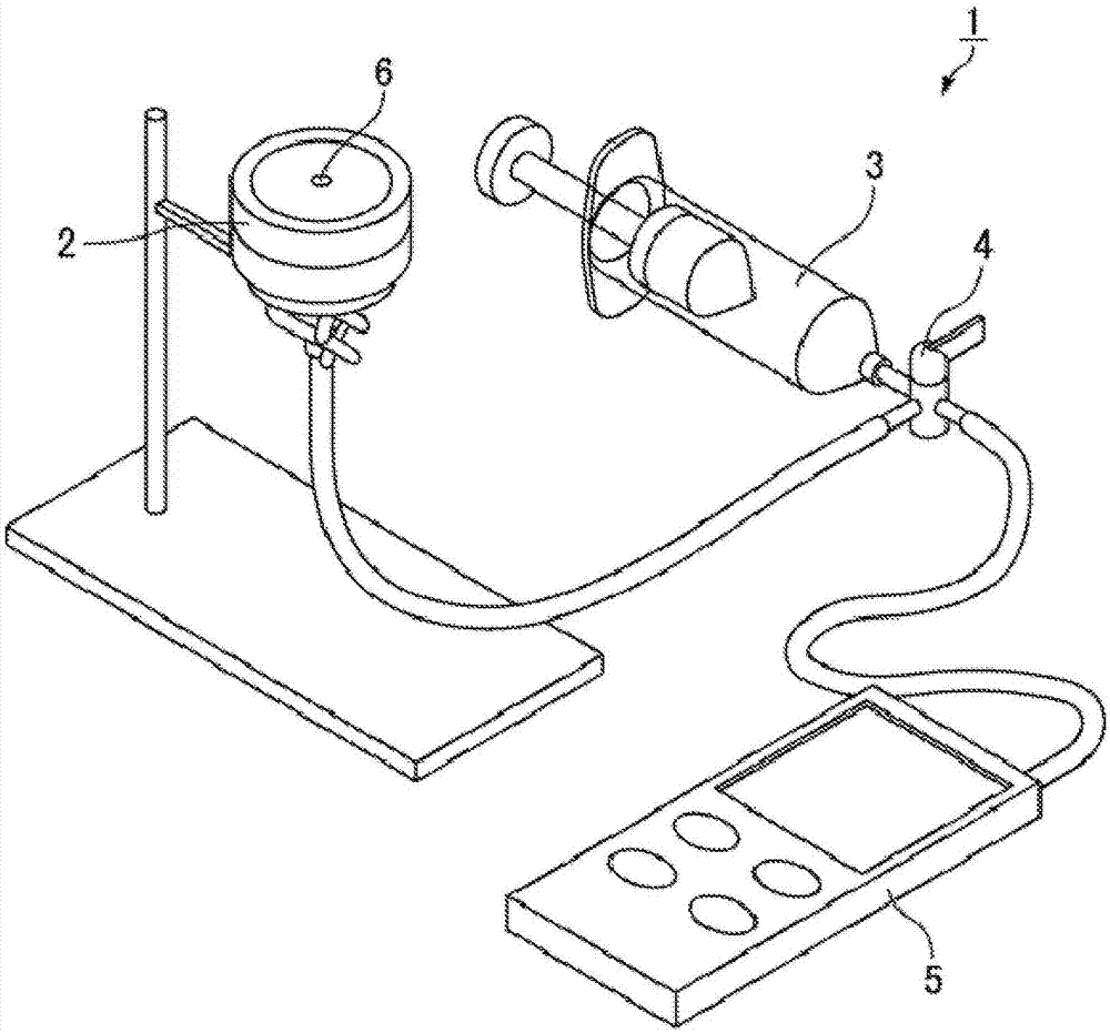
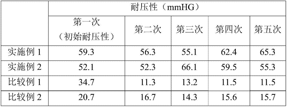
[0079] The prepared biological tissue reinforcement material was obtained in the same manner as in Example 1, except that the 200-μm-thick single-knit fabric made of 160-gauge cellulose yarn was used as a raw material to prepare the hydroxyethylated fiber fiber structure made of cellulose, and a combination of the three fiber structures made of hydroxyethylated cellulose/nonwoven made of polyglycolide/made of hydroxyethylated cellulose in the prescribed order The combination of the two fiber structures is laminated. Stress testing of biological tissue reinforcement materials. In the pressure test, the obtained test sample is placed in the center of the collagen membrane placed in the filter holder, so that the surface of the combined side of the three fibrous structures made of hydroxyethylated cellulose and the collagen membrane touch.
[0080] Table 1 shows the results of the stress test.

PUM

no PUM

Description & Claims & Application Information

We can also present the details of the Description, Claims and Application information to help users get a comprehensive understanding of the technical details of the patent, such as background art, summary of invention, brief description of drawings, description of embodiments, and other original content. On the other hand, users can also determine the specific scope of protection of the technology through the list of claims; as well as understand the changes in the life cycle of the technology with the presentation of the patent timeline. Login to view more.
Why Eureka
Try Eureka