
Fluorescence detection agent for p53 protein, and preparation method and application thereof

A fluorescent detection and protein technology, applied in the field of fluorescent detection agents for wild-type p53 protein, can solve the problems of high cost, complicated steps and the like, and achieve the effects of simple detection, simple operation and high sensitivity

Inactive Publication Date: 2018-02-02
CENT SOUTH UNIV
View PDF11 Cites 3 Cited by
  • Summary
  • Abstract
  • Description
  • Claims
  • Application Information

AI Technical Summary

Problems solved by technology

[0006] In order to solve the problems of cumbersome steps and high cost in the existing methods for detecting membrane proteins, the present invention provides a fluorescent detection agent for p53 protein with DNA functional modification, aiming at improving the sensitivity of fluorescent detection of p53 protein and reducing the experimental cost , to simplify the experimental steps

Method used


Image

  • Fluorescence detection agent for p53 protein, and preparation method and application thereof
  • Fluorescence detection agent for p53 protein, and preparation method and application thereof
  • Fluorescence detection agent for p53 protein, and preparation method and application thereof

Examples


Embodiment 1

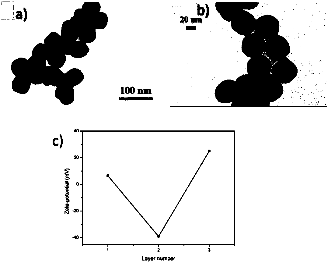
[0065] Step (1) Dextran-modified Fc 3 o 4 Preparation of NPs:

[0066] Fe 3 o 4 NPs were prepared by a hot solvent method. The specific process is as follows: 1.0g FeCl 3 ·6H 2 O was dissolved in 20 mL of ethylene glycol solution, and then 3.0 g of sodium acetate and 0.7 g of polyethylene glycol were added. After stirring for 30 min, the mixture was transferred to a polytetrafluoroethylene stainless steel autoclave, reacted at 200° C. for 8 h, and then cooled to room temperature. The precipitate was washed several times with ethanol and dried at 60°C to obtain Fe 3 o 4 NPs.

[0067] Next, PAA was used to modify the Fe by layer-by-layer self-assembly technique 3 o 4 NP s . The specific process is as follows: 1mL Fe 3 o 4 NPs (1mg mL -1 ) solution was added to 10mL PAA (1mg mL -1 ) solution, ultrasonically incubated for 15mim, collected with a magnet and washed several times with water, and then collected the PAA-coated Fe with a magnet 3 o 4 NPs(PAA / Fe 3 o ...

Embodiment 2

[0073] Step (1) dextran / PAA / Fe 3 o 4 The preparation of NPs: with the step 1 of embodiment 1

[0074] Step (2) DNA-2 / DNA-1 / dextran / PAA / Fe 3 o 4 The preparation of NPs: with the step 2 of embodiment 1

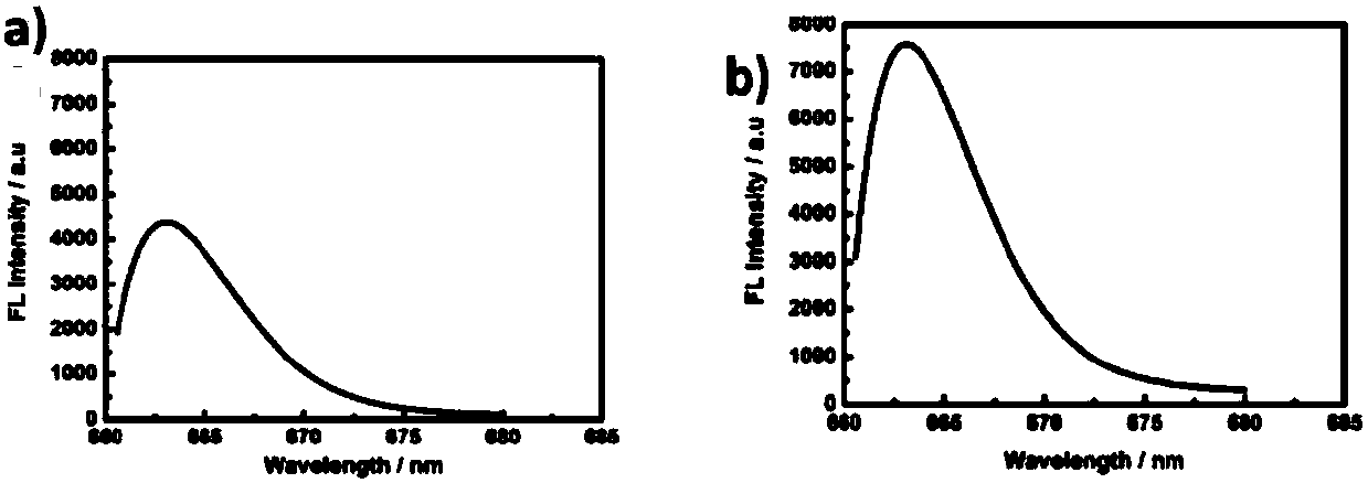
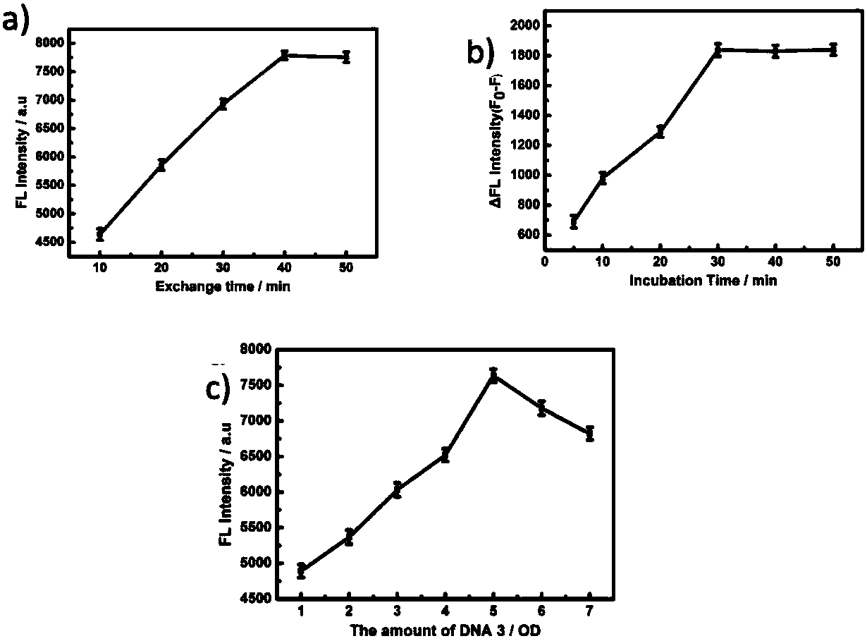
[0075] Step (3) Dilute p53 protein with PBS containing 20nM dithiothreitol (DTT), then add to DNA-2 / DNA-1 / dextran / PAA / Fe 3 o 4 In the NPs solution, after reacting for 30 minutes (incubation time), collect with a magnet and wash with PBS three times, and then collect nanoparticles that capture p53 protein with a magnet. Subsequently, Cy-5-labeled DNA-3 was added to replace DNA-2 (exchange reaction), and after heating at 37°C for 4 h (exchange reaction time), the nanoparticles (fluorescence detection agent) were collected and then redispersed in PBS solution, Test the fluorescence intensity of the fluorescence emission value at 663nm, such as figure 2 (b).

[0076] In order to explore the feasibility of using this strategy to detect p53 protein, 2nM p53 protein was added ...

Embodiment 3

[0078] Compared with Example 1, in order to investigate the influence of the DNA coverage on the surface of the magnetic nanoparticles on the detection sensitivity. Add DNA-1 with different concentration gradients from 1-7OD to 1mL dextran / PAA / Fe 3 o 4 NPs (1mg mL -1 ) solution, a total of 7 groups of magnetic nanoparticles modified by DNA-1 were obtained, and then DNA-2 corresponding to the amount of DNA-1 was added to each group to hybridize with DNA-1. Then, 1 nM p53 protein solution and the same amount of DNA-3 as DNA-1 were added to each group. The groups were then separated using a magnet, and the fluorescence intensity of the solution was measured. pass image 3 (c), inversely deduced that at 5OD, the DNA-1 load was the highest.


PUM


Abstract

The invention discloses a fluorescence detection agent for a p53 protein. The fluorescence detection agent comprises a magnetic nano-particle, double-stranded DNA, and a functional layer coating the surface of the magnetic nano-particle and connected with the double-stranded DNA; the double-stranded DNA is obtained through hybridizing DNA-1 single strand and DNA-2 single strand; the DNA-1 is a carboxyl oligonucleotide probe, and the sequence is COOH-(CH2)6-TTT TTT TCC GGG CAT GCC CGG CAT TGC-3'; and the DNA-2 is the partial complementary sequence of the DNA-1, and the sequence is 5'-GGG CAT GCC C-3'. The invention also discloses a preparation method of the fluorescence detection agent, and an application of the fluorescence detection agent in the detection of the wild p53 protein. The agent has the advantages of low preparation cost, realization of sensitive, rapid and accurate capture of the p53 protein, realization of ultra-sensitive, highly- selective and highly-accurate detection of the p53 protein, low detection limit, and realization of quantitative analysis and detection of the p53 protein in normal cells and cancer cell lysate, and can be used in clinical diagnosis and early detection of cancers.

Description

technical field [0001] The invention belongs to the technical field of p53 protein detection; in particular, it relates to a fluorescent detection agent for wild-type p53 protein. Background technique [0002] The incidence of cancer is increasing year by year, and it has become one of the major diseases threatening human health. It is estimated that cancer mortality is reduced by approximately 30% if cancer is detected and treated early. Therefore, early detection of cancer biomarkers has become a hotspot in cancer screening and prognosis research. As a tumor suppressor and transcription factor protein, p53 protein plays an important regulatory role in cell cycle, apoptosis and DNA repair. There are two types of p53 protein, wild-type and mutant. Mutations in the p53 gene increase to approximately 50% of all cancers, and in a wide variety of human cancers, mutations in this gene lead to accumulation of mutant p53 protein and reduction of wild-type p53 protein. Therefore...

Claims


Application Information

Patent Timeline
IPC IPC(8): G01N33/574G01N33/543
CPCG01N33/54326G01N33/57488G01N2333/4748
Inventor 邓留徐群芳刘珍军刘又年
Owner CENT SOUTH UNIV
Learn More