Magnetic resonance imaging contrast agent

A magnetic resonance contrast agent and magnetic resonance imaging technology, applied in the field of biological imaging, can solve the problems of short residence time, narrow imaging time window, and low relaxation rate, and achieve stable imaging time window, long imaging time window, biological good compatibility

Active Publication Date: 2020-12-25
JIANGSU PROVINCE INST OF TRADITIONAL CHINESE MEDICINE
View PDF4 Cites 0 Cited by
  • Summary
  • Abstract
  • Description
  • Claims
  • Application Information

AI Technical Summary

Problems solved by technology

At present, the commonly used contrast agents in clinical practice are small-molecule gadolinium-based paramagnetic contrast agents. This type of contrast agent has fast plasma clearance and a short residence time in the tissue, resulting in a narrow imaging time window; the relaxation rate is not high, and usually requires multiple administration, which brings a certain risk of toxicity; the most critical thing is that this type of contrast agent is a non-specific contrast agent, which cannot specifically image necrotic tissue
The magnetic resonance contrast agents used in the diagnosis of necrosis in the previous research are mainly porphyrin-based magnetic resonance contrast agents, but due to the phototoxicity of porphyrins, they have been discontinued, and there have been fewer and fewer studies in recent years.

Method used


Image

  • Magnetic resonance imaging contrast agent
  • Magnetic resonance imaging contrast agent
  • Magnetic resonance imaging contrast agent

Examples


Embodiment 1-1

[0043] Embodiment 1-1: the synthesis of formula (I) contrast agent

[0044] Add 200mg (0.70mmol) rhein and 148mg (0.77mmol) EDC to 100mL dichloromethane solution, stir at room temperature for 30min, then add 88mg (0.77mmol) NHS, react at room temperature for 24h until the reaction system is clear, add 100μLN -Boc-Ethylenediamine was added dropwise to the reaction solution, stirred at room temperature for 24 h, filtered to obtain a yellow solid, and washed with methanol to obtain pure intermediate 1.

[0045] 200 mg (0.47 mmol) of intermediate 1 was added to 15 ml of dichloromethane / trifluoroacetic acid (10:1), reacted at room temperature for 12 h, and the solvent was evaporated under reduced pressure to obtain intermediate 2.

[0046] Add 421mg (0.74mmol) DOTA-tris(tBu)ester, 280mg (0.74mmol) HBTU to 50mL acetonitrile, then add 100μL DIPEA, react at room temperature for 30min, then put 241mg (0.74mmol) intermediate 2 into the reaction system, Reaction 24h. The solvent was re...

Embodiment 1-2

[0050] Embodiment 1-2: the synthesis of formula (II) contrast agent

[0051] N-Boc-ethylenediamine in Example 1-1 was changed to N-Boc-butylenediamine, and the specific preparation method was the same as in Example 1-1 to obtain the magnetic resonance contrast agent of Ligand 2.

[0052] Ligand 2: 1 H NMR (300MHz, DMSO-d 6 ), δ(ppm): 11.81(s, 2H); 9.66(s, 1H); 8.18(s, 1H); 8.15(s, 1H); 7.90(s, 1H); 7.82~7.71(m, 2H) ;7.40(d, 1H, J=8.3Hz); 3.52~2.61(m, 28H); 1.57~1.55(m, 4H). 13 C NMR (75MHz, DMSO-d 6 ):δ:191.39,181.20,170.22,170.15,169.84,163.78,161.37,161.24,142.03,137.39,133.44,133.33,124.40,122.70,119.31,117.87,117.30,116.11,58.77,55.37,54.65,50.92,50.05, 49.94, 49.88, 37.93, 25.98, 25.65. ESI-MS m / z 777.2 for (M+K + -2H - ).

Embodiment 1-3

[0053] Embodiment 1-3: the synthesis of formula (III) contrast agent

[0054] N-Boc-ethylenediamine in Example 1-1 was changed to N-Boc-hexanediamine, and the specific preparation method was the same as in Example 1-1 to obtain the magnetic resonance contrast agent of Ligand 3.

[0055] Ligand 3: 1 H NMR (300MHz, DMSO-d 6 ), δ(ppm): 11.67(s, 2H); 9.14(s, 1H); 8.12(s, 2H); 7.83(s, 1H); 7.80~7.70(m, 2H); J=8.5Hz); 3.48~2.64(m, 28H); 1.56~1.32(m, 8H). 13 CNMR (75MHz, DMSO-d 6 ):δ:191.26,181.16,170.19,169.73,163.76,161.37,161.23,141.91,137.37,133.43,133.28,124.42,122.54,119.30,117.62,58.28,55.60,54.77,51.13,50.16,50.00,39.38,38.33, 28.74, 28.47, 25.95.ESI-MS m / z 805.3for ([M+K+2H] - ).


PUM


Abstract

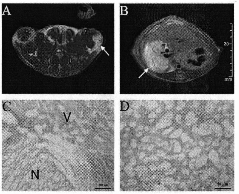
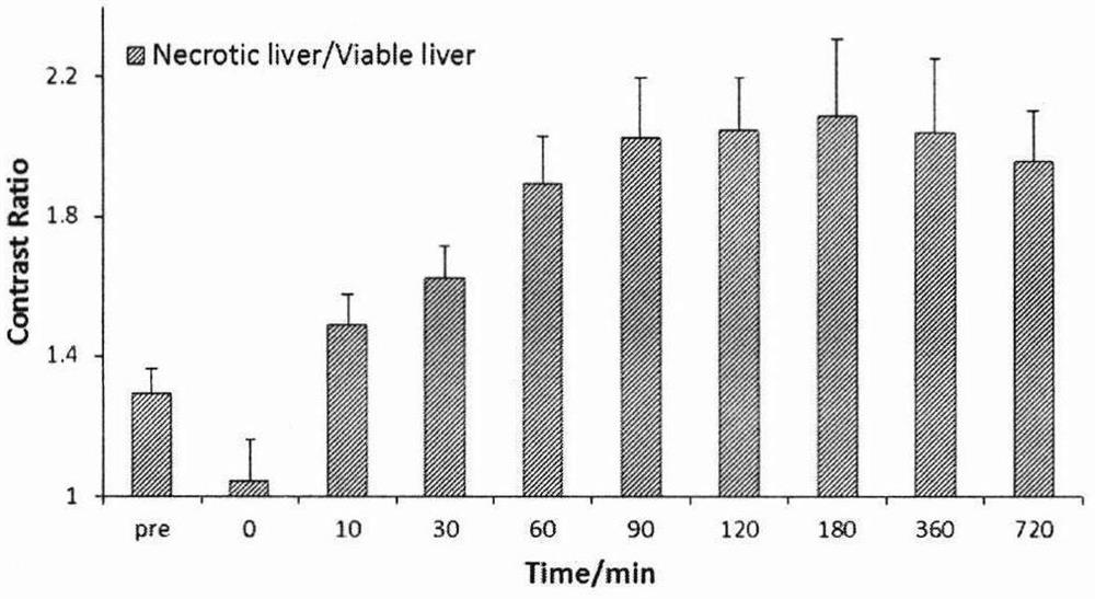
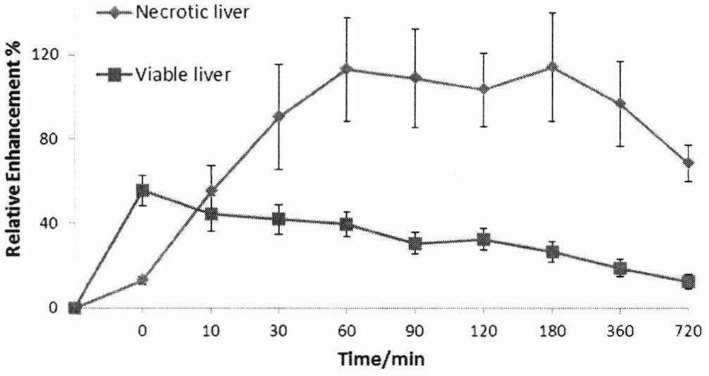
The invention relates to a class of magnetic resonance contrast agent, which is composed of three parts: a monoanthraquinone core, a connecting arm and an imaging part. This type of contrast agent can accumulate in necrotic liver, necrotic muscle, necrotic tissue in tumors, and infarcted myocardium, and can be used for the diagnosis of necrosis-related diseases, such as the evaluation of tumor treatment effects and the diagnosis of myocardial infarction. This type of contrast agent has a high relaxation rate; it stays in the necrotic tissue for a long time, and has a wide and stable imaging time window.

Description

[0001] field of invention [0002] The invention relates to a class of magnetic resonance contrast agents and belongs to the field of biological imaging. Background technique [0003] Necrosis is a form of cell death that manifests as a pathological state macroscopically. Tissue necrosis is a main feature of many diseases, such as cardiovascular disease, cancer, etc. Myocardial infarction, malignant tumors, and tumors will all form necrosis after non-surgical treatment. Therefore, imaging of necrotic tissue has great clinical application prospects for detecting necrosis-related diseases, such as being able to diagnose the disease, evaluate the prognosis of the disease, and monitor the effect of the disease on drugs. Treatment response, etc., so as to facilitate effective management of patients by doctors, guide doctors to quickly select the best treatment strategy to achieve individualized treatment, minimize side effects, and reduce medical expenses. [0004] Nowadays, mole...

Claims


Application Information

Patent Timeline
Patent Type & Authority Patents(China)
IPC IPC(8): A61K49/10
CPCA61K49/105A61K49/108
Inventor 张健卞丽高萌金乔梅蒋翠花张东建
Owner JIANGSU PROVINCE INST OF TRADITIONAL CHINESE MEDICINE
Why Eureka
Try Eureka