Method for adsorbing and separating extracellular vesicles including exosomes secreted to medium by cells based on anion exchange resin

A technology of resin exchange and adsorption separation, which is applied in the direction of embryonic cells, germ cells, animal cells, etc., can solve the problems that the concentration of exosomes cannot reach a high level, the price of ultracentrifuge tubes is expensive, and the time of researchers is wasted, so as to save costs and time, reduce high cost, and reduce the effect of operation difficulty

Active Publication Date: 2018-03-30
THE FIRST AFFILIATED HOSPITAL OF SUN YAT SEN UNIV
View PDF3 Cites 12 Cited by
  • Summary
  • Abstract
  • Description
  • Claims
  • Application Information

AI Technical Summary

Problems solved by technology

[0004] The most commonly used separation methods for extracellular vesicles and exosomes in the prior art are ultra-high-speed centrifugation or density gradient centrifugation. The price of centrifuge tubes is very expensive, and the centrifugation time is also very long, which consumes a lot of time for researchers; the use of ultracentrifuges requires individual tra

Method used


Image

  • Method for adsorbing and separating extracellular vesicles including exosomes secreted to medium by cells based on anion exchange resin
  • Method for adsorbing and separating extracellular vesicles including exosomes secreted to medium by cells based on anion exchange resin
  • Method for adsorbing and separating extracellular vesicles including exosomes secreted to medium by cells based on anion exchange resin

Examples


Embodiment 1

[0064] (1) A method for separating exosomes secreted from mesenchymal stem cells to the medium based on anion exchange resin adsorption

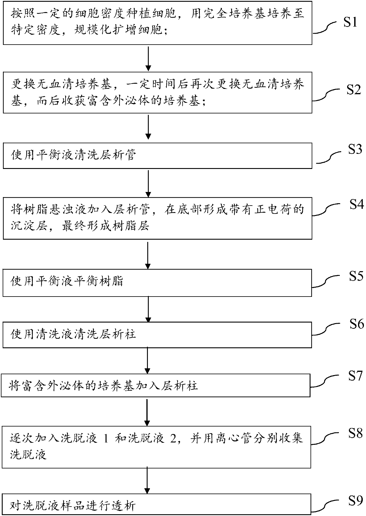
[0065] The method for separating extracellular vesicles secreted by mesenchymal stem cells into the medium based on anion exchange resin adsorption in this embodiment includes the following steps:

[0066] ①Use the complete medium of mesenchymal stem cells to cultivate mesenchymal stem cells from various sources in a massively expansive manner, with a planting density of 50-10000 / cm 2 , using the complete mesenchymal stem cell culture medium for 4-6 days, observe the cell growth density until 60-70%; the microscopic photos of the mesenchymal stem cells differentiated from urothelial-derived pluripotent induced stem cells are as follows image 3 shown;

[0067] ② When the cell density under the microscope reaches 60-70%, add 30-40mL / 175cm 2 serum-free medium. After 6 hours, harvest the first addition of serum-free medium and add a new 40...

Embodiment 2

[0092] (1) Macrophage function detection

[0093] 1.1 Experimental grouping (5 groups in total):

[0094] A. Normal control group: complete medium (DMEM+10%FBS, 300μL / well, the same below)

[0095] B. Positive control group: complete medium + 10ng / mL Ovalbumin (OVA)

[0096] C. Dexamethasone (DEX) treatment group: complete medium+10ng / mL OVA+1μM DEX

[0097] D. Exosome treatment group (5×10 8 / mL exosomes): complete medium+10ng / mL OVA+5×10 8 / mL exosomes

[0098] E. Exosome treatment group (2.5×10 9 / mL exosomes): complete medium+10ng / mL OVA+2.5×10 9 / mL exosomes

[0099] 1.2 Cell plate: resuscitate RAW 264.7 cells, divide the cells into 1×10 5 They were planted in 48-well culture plates, with 3 replicate wells in each group, and a total of 15 wells were planted. 300 μL of complete medium was added to each well, and placed in a cell culture incubator for overnight cultivation.

[0100] 1.3 Exosome anti-inflammatory function experiment: use 1.5mL EP tube to prepare 1mL...


PUM

PropertyMeasurementUnit
Diameteraaaaaaaaaa
Diameteraaaaaaaaaa

Abstract

The invention discloses a method for adsorbing and separating extracellular vesicles including exosomes secreted to a medium by cells based on an anion exchange resin, the extracellular vesicles including exosomes produced by the method, and application thereof. The method provided by the invention is simple and convenient to operate and only needs to use chromatographic tubes/columns and pipetteguns and other common laboratory consumables, does not require use of ultra centrifuges, saves cost and time; the obtained extracellular vesicles has high purity and large concentration, can be produced in a large scale, and is easy to be further applied clinically.

Description

technical field [0001] The present invention relates to a method for adsorbing extracellular vesicles including exosomes secreted by cells to a culture medium through an anion exchange resin, an application of exosomes produced by the method and extracellular vesicles including exosomes, and Treatment products. Background technique [0002] Exosomes are secreted by cells and belong to a kind of extracellular vesicles with a diameter of about 30-150nm. It has been found to play important physiological and pathological functions in various biological states and diseases. The exosome structure is similar to the cell membrane structure, consisting of a phospholipid bilayer with a thickness of about 5 nm, and its components mainly include ceramide, cholesterol, sphingolipids, and glycerophospholipids containing long saturated fatty chains. Exosomes are rich in various proteins on the surface and inside of vesicles, and contain important biological macromolecules such as multiple...

Claims


Application Information

Patent Timeline
IPC IPC(8): C12N5/0735C12N5/074C12N5/0775A61K35/28A61P29/00
CPCA61K35/28C12N5/0606C12N5/0663C12N5/0665C12N5/0667C12N5/0696C12N2500/38C12N2500/34C12N2500/32
Inventor 付清玲
Owner THE FIRST AFFILIATED HOSPITAL OF SUN YAT SEN UNIV
Why Eureka
Try Eureka