
Visual colorimetric sensor for detecting mercury ions

A visual colorimetric and sensor technology, applied in the field of analytical chemistry, can solve the problems of large background interference, high detection cost, complicated operation, etc., and achieve the effect of good reproducibility, good specificity and mild reaction

Active Publication Date: 2018-04-20
FUZHOU UNIV
View PDF6 Cites 9 Cited by
  • Summary
  • Abstract
  • Description
  • Claims
  • Application Information

AI Technical Summary

Problems solved by technology

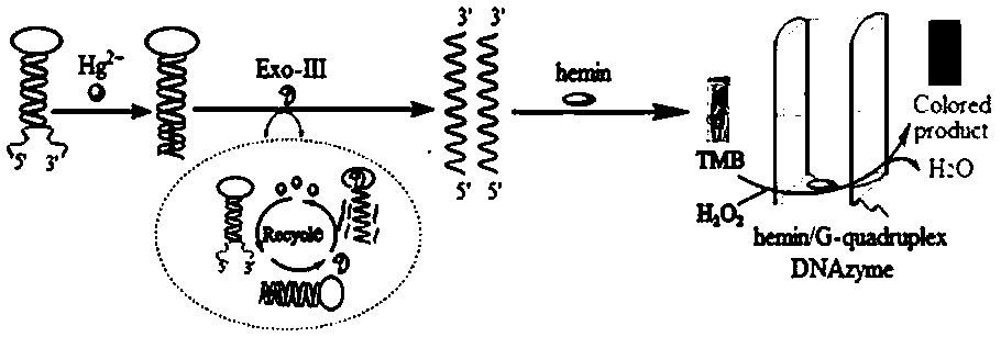
Design a H-DNA with a hairpin structure. In the presence of mercury ions, the stem part will be complementary and paired, triggering exonuclease III to cut it, and the resulting guanine-rich single-stranded DNA will be chlorinated In the presence of heme, DNA catalytic enzyme can be formed to catalyze 3,3',5,5'-tetramethylbenzidine (TMB) and H 2 o 2 By observing the obvious change of the color of the solution to judge whether the target analyte mercury ion exists and how much the content is, it can solve the problem of complicated operation, high requirements, high detection cost and high detection cost of traditional analysis and detection methods. Problems such as large background interference

Method used


Image

  • Visual colorimetric sensor for detecting mercury ions
  • Visual colorimetric sensor for detecting mercury ions
  • Visual colorimetric sensor for detecting mercury ions

Examples


Embodiment 1

[0034] 1. Preparation of H-DNA with hairpin structure:

[0035] (1) Prepare Tris-HCl buffer 1 (TB1, pH 7.4), TB1 consists of 20 mM Tris-HCl, 100 mM NaCl and 5mM MgCl 2 composition;

[0036] (2) Dissolve H-DNA in TB1, heat at 90°C for 10 minutes, and then cool to room temperature naturally.

[0037] 2. Preparation of hemin solution:

[0038] (1) Prepare Tris-HCl dilution buffer 2 (TB2, pH 7.4), TB2 consists of 300 mM Tris-HCl, 10mM TCEP, 1 M NaCl, 1mM EDTA and 1 mM MgCl 2 composition;

[0039] (2) First dissolve hemin in dimethyl sulfoxide (DMSO), and then dilute it to the required concentration with Tris-HCl buffer 2.

[0040] 3. Preparation of 3,3',5,5'-tetramethylbenzidine (TMB) solution:

[0041] (1) Prepare PBS buffer (pH 5.5), PBS buffer is made of 10 mM NaH 2 PO 4 , 10 mM Na 2 HPO 4 composition;

[0042] (2) First dissolve the TMB sample in pure ethanol, then add ultrapure water to volume, and dilute to the desired concentration with PBS buffer (pH 5.5).

Embodiment 2

[0044] Pretreatment of actual samples:

[0045] (1) After the environmental water sample was left to stand for a period of time, it was filtered with a 0.22 μm microporous membrane, and dilute nitric acid was added to the filtrate to make the HNO in the sample solution 3 The final concentration is 0.5%, mix well.

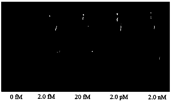
[0046] (2) Divide the tap water sample and the lake water sample into 3 parts, and number them: tap water No. 1, tap water No. 2, tap water No. 3 and lake water No. 1, lake water No. 2, and lake water No. Add standard Hg 2+ The solution was prepared with a spiked concentration of 1.0 fM, and the standard Hg was added to No. 2 tap water and No. 2 lake water. 2+ solution, prepared with a spiked concentration of 1.0 pM; add standard Hg to No. 3 tap water and No. 3 lake water 2+ solution, prepared with a spiked concentration of 1.0 nM.

Embodiment 3

[0048] Testing of actual samples:

[0049] (1) Take 45 μL of H-DNA with a concentration of 2.5 μM and 45 μL of water samples (or water sample spiked solutions with different concentrations), mix well, and add 10 μL of H-DNA with a concentration of 2.5 U / μL to them after 2 min. Exonuclease III (Exo-III), mix well and put it into a K30 economical dry thermostat, and react at 35 °C for 40 min;

[0050] (2) After the above reaction solution is cooled, add 100 μL of hemin (hemin) with a concentration of 7.75 μM to it, mix well and put it in the refrigerator (4°C) for 30 min;

[0051] (3) Add 100 μL of 5.0 mM 3,3',5,5'-tetramethylbenzidine (TMB) and 100 μL of 10.0 mM H 2 o 2 , and observe the color change of the solution after mixing for 1 min.


PUM


Abstract

The invention provides a visual colorimetric sensor for detecting mercury ions. An H-DNA (Hinge-Deoxyribonucleic Acid) with a special hairpin structure is designed; in the presence of the mercury ions, a 3' end and a 5' end of the H-DNA are complementary and paired through a T-Hg<2+>-T structure, so that a stem part of the H-DNA is completely complementary and exonuclease III is triggered to shearthe stem part, and then more and more single-stranded DNAs containing rich guanine are generated; released Hg<2+> also reacts with the H-DNA to trigger enzyme digestion reaction; the cycle is carriedout so that the H-DNA is continuously sheared and more and more single-stranded DNAs containing G are generated; in the presence of hematin chloride, a DNA catalyzing enzyme is formed, and 3,3',5,5'-tetramethylbenzidine is catalyzed to react with H2O2, so that a blue product is generated and the mercury ions are visibly detected. The method provided by the invention is simple to operate, low in cost and short in time, can be used for rapidly and visibly detecting the content of the mercury ions in an environment water sample and is hopefully applied to detection of mercury in the environmentwater sample.

Description

technical field [0001] The invention relates to a method for detecting mercury ions with a visual colorimetric sensor based on exonuclease III assisted target recycling and DNA catalytic enzymes to amplify signals, and belongs to the field of analytical chemistry. Background technique [0002] Mercury and mercury compounds are widely distributed and are one of the most harmful toxic heavy metals to humans and the environment in which they live. Due to the industrial use of mercury's toxicity and its application in industrial products such as preservatives, pesticides, and antibacterial agents, the probability of people's exposure to mercury and mercury compounds has greatly increased. Mercury ions can pose a huge threat to human health even at low concentrations. With the improvement of people's living standards and health awareness, more and more people begin to pay attention to the harm of mercury to human health. Studies have shown that mercury is a heavy metal element ...

Claims


Application Information

Patent Timeline
Patent Type & Authority Applications(China)
IPC IPC(8): G01N21/78
CPCG01N21/78
Inventor 许雪琴洪敏强李明玉曾碧花
Owner FUZHOU UNIV
Learn More