One-way amnion decellularization method

A decellularization and amniotic membrane technology, applied in the field of tissue engineering, can solve problems such as no solution is given, achieve the effect of simple operation, ensure stability, and facilitate large-scale production

Active Publication Date: 2018-05-18
上海睿泰生物科技股份有限公司
View PDF9 Cites 5 Cited by
  • Summary
  • Abstract
  • Description
  • Claims
  • Application Information

AI Technical Summary

Problems solved by technology

How to remove only the amniotic membrane epithelial cells and protect the amniotic m

Method used


Image

  • One-way amnion decellularization method
  • One-way amnion decellularization method
  • One-way amnion decellularization method

Examples


Embodiment 1

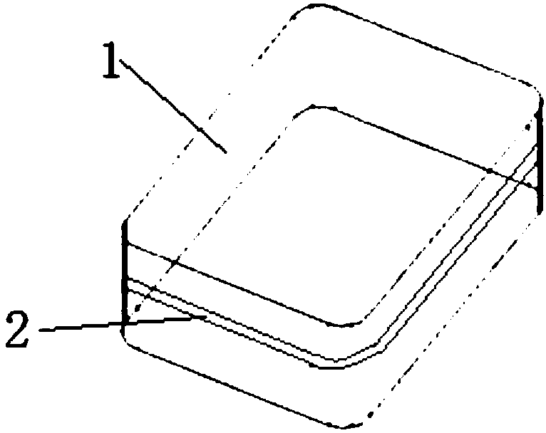
[0084] Take the placenta of healthy puerpera, strip the amniotic membrane from the placenta, wash it with 0.9% saline for several times in a biological safety cabinet, and use sterile medical tweezers to peel off the residual chorionic material and blood components. Spread the amniotic membrane on a sterile square one-way decellularization device (20x20x2cm, figure 1 as shown, figure 11 refers to the plane, 2 refers to the groove on the side) (the stroma layer is down), and use a sterile elastic medical latex tube to wrap the edge of the amniotic membrane on the groove of the device and fix it, use sterile medical scissors along the groove Unfixed amniotic membrane was cut off. Put the fixed amnion into TrypLE TM In the Select Enzyme solution, shake at 37°C for 2 hours. After shaking and washing with sterile normal saline for 5 times, the fixed amniotic membrane was put into Super Nuclease solution and treated with shaking at 37°C for 3 hours. Finally, the human amniotic ...

Embodiment 2

[0092] Take the placenta of healthy puerpera, strip the amniotic membrane from the placenta, wash it with 0.9% saline for several times in a biological safety cabinet, and use sterile medical tweezers to peel off the residual chorionic material and blood components. Spread the amniotic membrane on a sterile cylindrical one-way decellularization device (15 cm in diameter and 2 cm in height) (with the stroma layer facing down), and wrap the edge of the amnion on the groove of the device with a sterile elastic medical rubber band to fix it. Cut off the unfixed amniotic membrane along the groove with sterile medical scissors. Put the fixed amniotic membrane into the neutral protease solution, and shake at 37°C for 30 minutes. After shaking and washing with sterile normal saline for 5 times, the fixed amnion was put into Super Nuclease solution and treated with shaking at 37°C for 1 hour. Finally, the human amniotic membrane extracellular matrix scaffold material was obtained by s...

Embodiment 3

[0097] Take the placenta of healthy puerpera, strip the amniotic membrane from the placenta, wash it with 0.9% saline for several times in a biological safety cabinet, and use sterile medical tweezers to peel off the residual chorionic material and blood components. Spread the amniotic membrane on the sterile rectangular one-way decellularization device (20x15x3cm) (stromal layer down), wrap the edge of the amniotic membrane on the groove of the device with a sterile elastic medical rubber band and fix it with sterile medical scissors Cut off the unfixed amniotic membrane at the groove. Put the fixed amniotic membrane into CHAPS solution, shake at 37°C for 12h. After shaking and washing with sterile normal saline for 5 times, the fixed amniotic membrane was put into Benzonase Nuclease solution and treated with shaking at 37°C for 8 hours. Finally, the human amniotic membrane extracellular matrix scaffold material was obtained by shaking and washing with sterile normal saline ...


PUM

PropertyMeasurementUnit
Diameteraaaaaaaaaa

Abstract

The invention relates to a one-way amnion decellularization method. The method comprises the following steps that a, a fresh amnion is stripped and cleaned; b, the amnion obtained in step a is laid and fixed to a supporting body with a plane; c, the amnion processed in step b is placed into a decellularization solution for treatment; d, the amnion treated in step c is washed and then placed into anuclease solution for treatment; e, the amnion treated in step d is washed, and then a human decellularized amnion is obtained. Compared with the prior art, the method is combined with structure characteristics of the amnion, and a one-way decellularization method is used for protecting a matrix layer of the amnion; the amnion is laid and expanded, an epithelial surface fully makes contact with the decellularization solution, so that epithelial cells of the amnion are thoroughly removed, and a very small number of cells of the matrix layer are removed by making contact with a small quantity of decellularization solution due to the permeability of tissue. The decellularization method is easy and convenient to operate, the stability of the process is guaranteed, and large-scale production is facilitated.

Description

technical field [0001] The invention relates to the technical field of tissue engineering, in particular to a one-way amniotic membrane decellularization method. Background technique [0002] Amniotic membrane, that is, the human placental mucosa, is the innermost layer of the fetal membrane, which is closely attached to the chorion. The normal thickness of the amniotic membrane is 0.02-0.5mm. It is a smooth, translucent film with certain toughness. It has no blood vessels, nerves and lymphatic vessels. It consists of a single epithelial cell layer, a dense basement membrane, and an avascular stroma. Amniotic membrane stroma contains a large amount of collagen, fibronectin and laminin, etc. Because of its growth factors, extremely low immunogenicity, antibacterial, anti-inflammatory, anti-fibrosis, and inhibition of neovascularization, it is widely used in the medical field. With a history of more than 100 years of application, its main uses include ocular surface disease r...

Claims


Application Information

Patent Timeline
IPC IPC(8): C12N5/073A61L27/36
CPCA61L27/3604A61L27/3687A61L2430/40C12N5/0605
Inventor 董教明吴复跃何振东
Owner 上海睿泰生物科技股份有限公司
Why Eureka
Try Eureka