Medical ultrasonic coupling patch preparation method
An ultrasonic coupling and patch technology, applied in the medical field, achieves the effects of easy use, clear ultrasonic images, and excellent sound guiding performance
- Summary
- Abstract
- Description
- Claims
- Application Information
AI Technical Summary
Problems solved by technology
Method used
Image
Examples
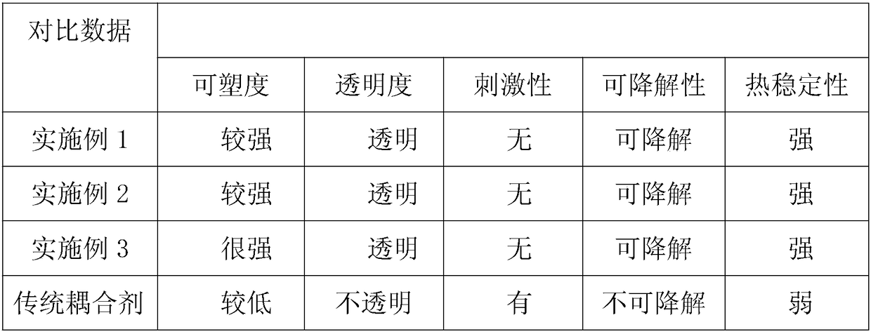
Embodiment 1
[0023] A method for preparing a medical ultrasonic coupling patch includes the following components: purified water, reactive monomers, reactive crosslinking agents, preservatives and initiators. In terms of weight ratio, the proportions of each component are: purified water 80%, reactive monomer 15%, reactive crosslinking agent 4%, preservative 0.05%, initiator 0.95%, including the following preparation steps:
[0024] Step 1. Slowly add alginic acid to purified water, stir until completely dissolved, and leave it aside;
[0025] Step 2. Prepare acrylamide and add it to purified water, stir until it is completely dissolved, and leave aside;
[0026] Step 3. Prepare ammonia persulfate and add it to purified water, stir until it is completely dissolved, and leave it aside;
[0027] Step 4. Prepare the purified water to which paraben is added, stir until completely dissolved, and leave it aside;
[0028] Step 5. Stir the above-mentioned mixture to be reacted evenly and then inject it int...
Embodiment 2
[0036] A preparation method of medical ultrasonic coupling patch includes purified water, reactive monomer, reactive cross-linking agent, preservative and initiator. In terms of weight ratio, the proportions of each component are: 85% of purified water, 12% of reactive monomer, 2% of reactive crosslinking agent, 0.02% of preservative, and 0.98% of initiator, including the following preparation steps:
[0037] Step 1. Slowly add methyl methacrylate alcohol solution to purified water, stir until completely dissolved, and leave aside;
[0038] Step 2. Prepare acrylamide and add it to purified water, stir until it is completely dissolved, and leave aside;
[0039] Step 3. Prepare azobisisobuimidazoline hydrochloride and add it to purified water, stir until completely dissolved, and leave it aside;
[0040] Step 4. Prepare the purified water to which paraben is added, stir until completely dissolved, and leave it aside;
[0041] Step 5. Stir the above-mentioned mixture to be reacted evenly ...
Embodiment 3
[0049] A preparation method of medical ultrasonic coupling patch includes purified water, reactive monomer, reactive crosslinking agent, preservative and initiator. By weight, the proportions of each component are: purified water 82%, reactive monomer 13%, reactive crosslinking agent 3.5%, preservative 0.03%, initiator 1.42%, including the following preparation steps:
[0050] Step 1. Slowly add methyl methacrylate alcohol solution to purified water, stir until completely dissolved, and leave aside;
[0051] Step 2. Prepare ethylene glycol dimethacrylate and add it to purified water, stir until it is completely dissolved, and leave it aside;
[0052] Step 3. Prepare azobisisobutylamidine hydrochloride and add it to purified water, stir until completely dissolved, and leave aside;
[0053] Step 4. Prepare purified water to which dehydroacetic acid and sodium salt are added, stir until completely dissolved, and leave aside;
[0054] Step 5. Stir the above-mentioned mixture to be reacted ...
PUM
 Abstract
Description
Claims
Application Information
 - R&D
- Intellectual Property
- Life Sciences
- Materials
- Tech Scout
- Unparalleled Data Quality
- Higher Quality Content
- 60% Fewer Hallucinations
Browse by: Latest US Patents, China's latest patents, Technical Efficacy Thesaurus, Application Domain, Technology Topic, Popular Technical Reports.
© 2025 PatSnap. All rights reserved.Legal|Privacy policy|Modern Slavery Act Transparency Statement|Sitemap|About US| Contact US: help@patsnap.com

