
Single-cell genome copy number variation detection method and kit

A detection method and single-cell technology, applied in the biological field, can solve problems such as false positives, false positive results, and different amounts of sequencing data

Inactive Publication Date: 2018-08-17
CAPITALBIO CORP
View PDF4 Cites 6 Cited by
  • Summary
  • Abstract
  • Description
  • Claims
  • Application Information

AI Technical Summary

Problems solved by technology

The selection of window length and CNV in the prior art often leads to false positive results
The selection of window length in existing technologies is usually for specific research purposes. The amount of sequencing data is different, the resolution requirements for detecting CNV are different, and the optimal window length required is also different. The same data using too large a window may lead to false negatives As a result, a window that is too small can lead to false positive results

Method used


Image

  • Single-cell genome copy number variation detection method and kit
  • Single-cell genome copy number variation detection method and kit
  • Single-cell genome copy number variation detection method and kit

Examples


Embodiment 1

[0126] 1.1 Single cell picking

[0127] Pick single amniocytes using a mouth pipette or flow cytometer.

[0128] 1.2 Single cell expansion

[0129] 1.2.1 Cell Lysis

[0130] 1.2.1.1 According to the number of reactions, mix Cell Lysis Buffer and Cell Lysis Enzyme to prepare the cell lysis mixture.

[0131] Table 1

[0132] Cell Lysis Mixture

volume

Cell Lysis Buffer

2.5μL

Cell Lysis Enzymes

0.05μL

total capacity

2.55 μL

[0133] 1.2.1.2 Collect single cells in a PCR tube containing 2.5 μL of cell lysis mixture.

[0134] 1.2.1.3 Incubate the sample in a preheated PCR machine under the following conditions:

[0135] Table 2

[0136]

[0137] 1.2.1.4 After the procedure ends, briefly centrifuge to collect the reaction solution.

[0138] 1.2.2 Pre-amplification

[0139] 1.2.2.1 Mix Pre-Amp Buffer and Pre-Amp Enzyme Mix to prepare the pre-amplification mixture.

[0140] table 3

[0141] preamplification mix

...

Embodiment 2~4

[0190] 1.1 Single cell picking

[0191] Pick single amniocytes using a mouth pipette or flow cytometer.

[0192] 1.2 Single cell expansion

[0193] 1.2.1 Cell Lysis

[0194] 1.2.1.1 According to the number of reactions, mix Cell Lysis Buffer and Cell Lysis Enzyme to prepare the cell lysis mixture.

[0195] Table 10

[0196] Cell Lysis Mixture

volume

Cell Lysis Buffer

2.5μL

Cell Lysis Enzymes

0.05μL

total capacity

2.55 μL

[0197] 1.2.1.2 Collect single cells in a PCR tube containing 2.5 μL of cell lysis mixture.

[0198] 1.2.1.3 Incubate the sample in a preheated PCR machine under the following conditions:

[0199] Table 11

[0200]

[0201] 1.2.1.4 After the procedure ends, briefly centrifuge to collect the reaction solution.

[0202] 1.2.2 Pre-amplification

[0203] 1.2.2.1 Mix Pre-Amp Buffer and Pre-Amp Enzyme Mix to prepare the pre-amplification mixture.

[0204] Table 12

[0205] preamplification ...

Embodiment 5

[0288] Embodiment 5 data analysis

[0289] Table 19

[0290]

[0291]

[0292]

[0293] Analysis conclusion:

[0294] From the above analysis results, it can be seen that the data quality Q30 and uniformity of the method in this case are significantly higher than that of the comparison method, and a smaller data volume (0.05Gb) and lower coverage (1.2%) are achieved. Chromosomal microamplification and microdeletion detection.


PUM


Abstract

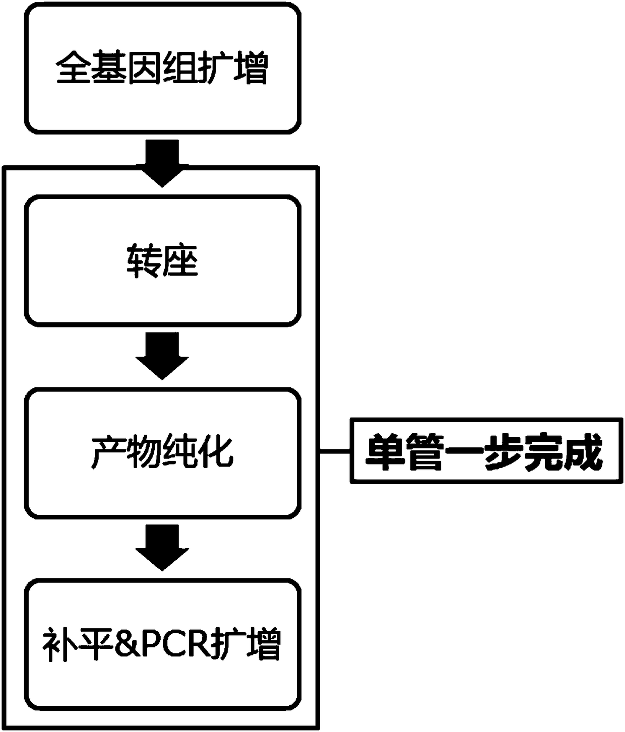
The invention relates to the technical field of biology, in particular to a combination, a kit and application of the kit. The application includes a solution of single-cell genome copy variation detection, comprising single cell amplification, library construction, high-throughput sequencing and information analysis; library construction based on transposase is applied to single cell sequencing;the use of single-tube one-step library construction process helps relieve operational complexity, thus avoiding pollution. In addition, the invention also provides optimal window length according tothe requirement on CNV (copy number variation) lowest detection resolution; run test is performed to count distribution differences, relative to coverage, of CNV area and normal area sequencing data;significance P value of each candidate CNV is output in final results; detection accuracy is effectively improved.

Description

technical field [0001] The invention relates to the field of biotechnology, in particular to a rapid detection method and kit for single-cell genome copy number variation. Background technique [0002] Single-cell whole-genome sequencing technology is a new technology for amplifying and sequencing the whole genome at the single-cell level. The principle is to amplify a small amount of whole-genome DNA from a single isolated cell, obtain a complete genome with high coverage, and perform high-throughput sequencing, which can be used to detect single-cell genome copy number variation (including chromosomal aneuploidy and chromosomal aneuploidy). Micro-amplification and micro-deletion), revealing individual differences and cell evolution relationships in cell populations. [0003] For single-cell whole-genome sequencing, the library construction process of single-cell amplification products involves DNA fragmentation, end filling, adenylation, adapter ligation, and PCR amplific...

Claims


Application Information

Patent Timeline
IPC IPC(8): C12Q1/6869C12Q1/6858C40B50/06
CPCC12Q1/6858C12Q1/6869C40B50/06C12Q2535/122C12Q2525/191
Inventor 郭弘妍田婕邓莉莉邢婉丽程京张怡然
Owner CAPITALBIO CORP
Learn More