Preparation method and application of rare earth cerium-quantum dot coordination polymer double-emission fluorescent probe

A dual-emission fluorescence and coordination polymer technology, applied in the field of fluorescence sensing, achieves the effects of good selectivity, improved accuracy, and reduced detection limit

Inactive Publication Date: 2018-09-04
NANCHANG UNIV
View PDF4 Cites 3 Cited by
  • Summary
  • Abstract
  • Description
  • Claims
  • Application Information

AI Technical Summary

Problems solved by technology

The reference in this internal standard ratiometric fluorescent probe has a constant fluorescent signal and is insensitive to analyte recognition

Method used


Image

  • Preparation method and application of rare earth cerium-quantum dot coordination polymer double-emission fluorescent probe
  • Preparation method and application of rare earth cerium-quantum dot coordination polymer double-emission fluorescent probe
  • Preparation method and application of rare earth cerium-quantum dot coordination polymer double-emission fluorescent probe

Examples


Embodiment 1

[0032] Preparation of rare earth cerium-quantum dot coordination polymer dual emission fluorescent probe

[0033] (1) Add 30 μL of 10 mM adenosine triphosphate (ATP) to 300 μL of 50 mM pH 7.4 Tris-HCl buffer solution, mix well, and then add 500 μL of 40 nM carboxy quantum dots (QDs ) and 200μL 4mM Ce(NO 3 ) 3 Solution, stirred slowly at room temperature for 2 minutes, forming a white turbid solution;

[0034] (2) Centrifuge the white turbid liquid at 16500rpm for 10min to obtain a white precipitate, wash it three times with ultrapure water, resuspend the white precipitate in 1mL of ultrapure water, and make a rare earth cerium-quantum dot coordination polymer double emission Fluorescent probes (hereinafter referred to as Ce-QDs CPNs).

Embodiment 2

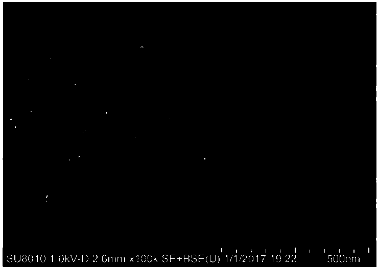
[0036] Microscanning of Ce-QDs CPNs

[0037] The morphology of the Ce-QDs CPNs prepared in Example 1 was characterized by scanning electron microscope (SEM), the results are as follows figure 1 shown.

[0038] From figure 1 It can be seen that the Ce-QDs CPNs prepared by the method of the present invention are composed of spherical particles with a diameter of about 10 nm to 30 nm. This is mainly due to the fact that the ATP molecule contains a phosphate group with multi-coordinating ability, which converts the rare earth Ce 3+ Rare earth coordination polymers were formed through coordination with carboxylated quantum dots (QDs) in Tris-HCl buffer solution.

Embodiment 3

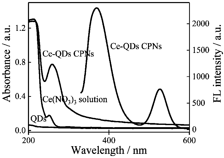
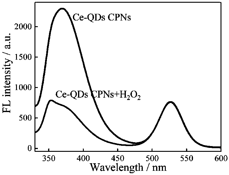
[0040] QDs, Ce(NO 3 ) 3 , UV spectra of Ce-QDs CPNs and fluorescence spectra of Ce-QDs CPNs.

[0041] 2nM QDs solution, 0.08mM Ce(NO 3 ) 3 ·6H 2 UV-Vis absorption spectra of O solution and 0.08 mM Ce-QDs CPNs solution. The fluorescence emission spectrum of the Ce-QDs CPNs solution at the excitation wavelength of 310 nm was measured by a fluorescence spectrometer. The result is as figure 2 shown.

[0042] From figure 2 It can be seen from the UV-visible absorption spectrum that Ce(NO 3 ) 3 ·6H 2 The characteristic ultraviolet absorption peak of O solution is 252nm, QDs has almost no ultraviolet absorption peak, and Ce-QDs CPNs has a strong and broad absorption peak at 260nm. Under the excitation wavelength of 310nm, the Ce(III) and QDs fluorescence peaks of Ce-QDs CPNs appeared at 369nm and 525nm, respectively. Ce by spectrometry 3+ , ATP and QDs self-assembly behavior in Tris-HCl buffer solution was characterized. Ce-QDs CPNs at 1710(νP-OH), 1480(νN7-C8), 1258(...


PUM


Abstract

The invention provides a preparation method of a rare earth cerium-quantum dot coordination polymer double-emission fluorescent probe. The method comprises the following steps that (1) triphosadenineis added into a tromethamine hydrochloride buffer solution; after the uniform mixing, carboxyl quantum dots and a Ce(NO3)3 solution are added; slow stirring and uniform mixing are performed at room temperature; a white turbid solution is formed; (2) the white turbid solution obtained in the first step is centrifugalized at a certain rotating speed for certain time; white precipitates are obtained;ultrapure water is used for cleaning for three times; the white precipitates are resuspended in 1mL of ultrapure water; the rare earth cerium-quantum dot coordination polymer double-emission fluorescent probe is prepared. The prepared double-emission fluorescent probe can be used for sensitive detection of hydrogen peroxide and glucose.

Description

technical field [0001] The invention relates to the technical field of fluorescence sensing, in particular to a preparation method and application of a rare earth cerium-quantum dot coordination polymer dual-emission fluorescent probe. Background technique [0002] Hydrogen peroxide (H 2 o 2 ) as an important chemical and a by-product of many enzymatic reactions has been playing an important role in the fields of medicine, pharmacy, food safety, environmental analysis and industrial production. Therefore, fast and accurate quantitative detection of H 2 o 2 is of great significance. So far, scientists have developed many detection methods for H 2 o 2 methods, such as electrochemical methods, fluorescence methods, colorimetric methods, and chromatography. Among them, fluorescence methods have received more and more attention due to their advantages such as small sample size, convenient operation, and diverse designs. Chen et al established the H 2 o 2 Fluorescence det...

Claims


Application Information

Patent Timeline
IPC IPC(8): C08G83/00C09K11/06G01N21/64
CPCC08G83/008C09K11/06C09K2211/182G01N21/6402
Inventor 梁汝萍杨佳乐邱建丁
Owner NANCHANG UNIV
Why Eureka
Try Eureka