
Microsphere for double protection of antibody drug and intravitreal injection, and preparation method thereof

A double-protection, anti-antibody technology, applied in the field of medicine, can solve problems such as the reduction of drug loading and encapsulation efficiency, antibody denaturation and aggregation, and large molecular weight of drugs, so as to improve denaturation and aggregation, reduce irritation, and improve patient compliance Effect

Active Publication Date: 2018-12-07
南京锐利施生物技术有限公司
View PDF2 Cites 3 Cited by
  • Summary
  • Abstract
  • Description
  • Claims
  • Application Information

AI Technical Summary

Problems solved by technology

However, due to the large molecular weight and fragile structure of the drug, the organic phase / water phase interface generated during the preparation process is likely to cause denaturation and aggregation of antibodies, resulting in a decrease in drug activity.
Moreover, the drug in the inner water phase is easy to diffuse into the outer water phase due to hydrophilicity, resulting in lower drug loading and encapsulation efficiency.

Method used


Image

  • Microsphere for double protection of antibody drug and intravitreal injection, and preparation method thereof
  • Microsphere for double protection of antibody drug and intravitreal injection, and preparation method thereof
  • Microsphere for double protection of antibody drug and intravitreal injection, and preparation method thereof

Examples


Embodiment 1

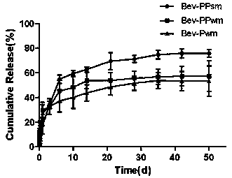
[0029] Note: Bev-PPsm in the experimental group is PLGA / PCADK microspheres loaded with bevacizumab prepared by S / O / W method; Bev-PPwm in the control group is loaded with bevacizumab prepared by W / O / W PLGA / PCADK microspheres of vacizumab; the control group Bev-Pwm is PLGA microspheres loaded with bevacizumab prepared by W / O / W method;

[0030] Bev-PPsm Prescription

[0031] Phase S: ​​bevacizumab-dextran microparticles (1:3), 50mg;

[0032] Phase O: PLGA 7525 4A, 80 mg; PCADK, 20 mg; Dichloromethane, 2 mL;

[0033] Phase W: 1.0% PVA aqueous solution, 10 mL;

[0034] Preparation process of Bev-PPsm

[0035] 1) Dissolve bevacizumab, dextran and PEG (1: 3: 40) in deionized water, vortex and then freeze-dry; the powder obtained after freeze-drying is washed by centrifugation with dichloromethane to remove the PEG dispersed phase, Vacuum drying to obtain bevacizumab-dextran microparticles;

[0036] 2) Dissolve PLGA and PCADK in dichloromethane, add the prepared particles, and ul...

Embodiment 2

[0050] Note: Afl-PPsm in the experimental group is the PLGA / PCADK microspheres loaded with aflibercept prepared by the S / O / W method; Afl-PPwm in the control group is prepared by the W / O / W method PLGA / PCADK microspheres of Aflibercept; the control group Afl-Pwm is PLGA microspheres loaded with Aflibercept prepared by W / O / W method;

[0051] Afl-PPsm prescription

[0052] Phase S: ​​Aflibercept-dextran microparticles (1:4), 40mg;

[0053] Phase O: PLGA 7525 4A, 70mg; PCADK, 30mg; Dichloromethane, 2 mL;

[0054] Phase W: 2.0% PVA aqueous solution, 15 mL;

[0055] Preparation process of Afl-PPsm

[0056] 1) Dissolve aflibercept, dextran and PEG (1:4:40) in deionized water, vortex and then freeze-dry; the powder obtained after freeze-drying is washed by centrifugation with dichloromethane to remove the PEG dispersed phase, Vacuum drying to obtain aflibercept-dextran microparticles;

[0057] 2) Dissolve PLGA and PCADK in dichloromethane, add the prepared particles, and ultrasonica...

Embodiment 3

[0071] Note: The experimental group Luc-PPsm is the PLGA / PCADK microspheres loaded with ranibizumab prepared by the S / O / W method; the control group Luc-PPwm is the ranibizumab-loaded microspheres prepared by the W / O / W method PLGA / PCADK microspheres of zizumab; the control group Luc-Pwm is PLGA microspheres loaded with ranibizumab prepared by W / O / W method;

[0072] Luc-PPsm Prescription

[0073] Phase S: ​​ranibizumab-dextran microparticles (1:5), 48mg;

[0074] Phase O: PLGA 7525 5A, 80mg; PCADK, 20mg; Dichloromethane, 2 mL;

[0075] Phase W: 1.0% PVA aqueous solution, 20 mL;

[0076] Preparation process of Luc-PPsm

[0077] 1) Weigh ranibizumab, dextran and PEG (1:5:40) and dissolve in deionized water, vortex and then freeze-dry; the powder obtained after freeze-drying is centrifuged with dichloromethane to remove the PEG dispersed phase, Vacuum drying to obtain ranibizumab-dextran microparticles;

[0078] 2) Dissolve PLGA and PCADK in dichloromethane, add the prepared p...


PUM

PropertyMeasurementUnit
particle diameteraaaaaaaaaa

Abstract

The invention provides a microsphere for double protection of antibody drug and intravitreal injection, and a preparation method thereof. Particles of a monoclonal antibody drug are prepared by adopting a method of water phase-water phase emulsification, polylactic acid-glycolic acid copolymer and polyketal are utilized as carrier materials, and a sustained release microsphere that wraps and carries the particles of a dextran-monoclonal antibody drug is prepared by employing an emulsion solvent volatilization method of water phase-oil phase-solid phase. Denaturation and aggregation of antibodies are caused by an acidic microenvironment of polylactic acid-glycolic acid in an organic phase / aqueous phase interface and a degradation process, the drug loading capacity of the microsphere is increased and a burst release phenomenon during a releasing process of the microsphere is reduced, and double protection on the monoclonal antibody drug of the microsphere is achieved; the polyketal enables the drug loading capacity of the microsphere to be increased, enables the stimulation of an acidic degradation product of the polylactic acid and glycolic acid on eye environment to be reduce, andenables side effects such as endophthalmitis to be reduced; and the microsphere can release the monoclonal antibody for not less than 28 days in the in vitro environment and for not less than 2 monthsin the eye, and so, the frequency of administration can be reduced, pain of patients and economic burden are reduced, and the compliance of patients is improved.

Description

technical field [0001] The invention relates to a microsphere injected into the vitreous with double protection against antibody drugs. The invention further provides a preparation method thereof, which belongs to the technical field of medicine. Background technique [0002] Retinal diseases have become the second leading cause of blindness after cataract in my country, among which age-related macular degeneration (AMD) is recognized as one of the most difficult eye diseases to treat, and choroidal neovascularization (CNV) is the most important cause of vision loss. reason. Among them, vascular endothelial growth factor (VEGF) and its receptor play an important role in the proliferation and repair of pathological neovascularization. Abnormal angiogenesis causes vascular leakage in ocular vascular disease. Therefore, anti-VEGF drugs become the key to the treatment of wet age-related macular degeneration. [0003] Aflibercept (Eylea®) is the world's first fully humanized fu...

Claims


Application Information

Patent Timeline
Patent Type & Authority Applications(China)
IPC IPC(8): A61K9/52A61K47/34A61K39/395A61P27/02
CPCA61K9/0019A61K9/0048A61K9/5031A61K9/5089A61K39/395A61P27/02
Inventor 李又欣刘佳欣孙凤英李爽滕乐生
Owner 南京锐利施生物技术有限公司
Learn More