LED-excited short wavelength infrared fluorescent microscopic imaging system

A microscopic imaging and short-wave infrared technology, which is applied in fluorescence/phosphorescence, material excitation analysis, material analysis by optical means, etc., can solve the problems of fluorescence microscopic imaging, etc., and achieve small damage to biological tissue, broad development and improvement. Application prospect, the effect of strong signal

Inactive Publication Date: 2018-12-11
ZHEJIANG UNIV
View PDF4 Cites 11 Cited by
  • Summary
  • Abstract
  • Description
  • Claims
  • Application Information

AI Technical Summary

Problems solved by technology

There are some formed fluorescence microscopy imaging systems at home and abroad, but there i

Method used


Image

  • LED-excited short wavelength infrared fluorescent microscopic imaging system
  • LED-excited short wavelength infrared fluorescent microscopic imaging system

Examples


Example Embodiment

[0018] The present invention will be further described below in conjunction with the drawings.

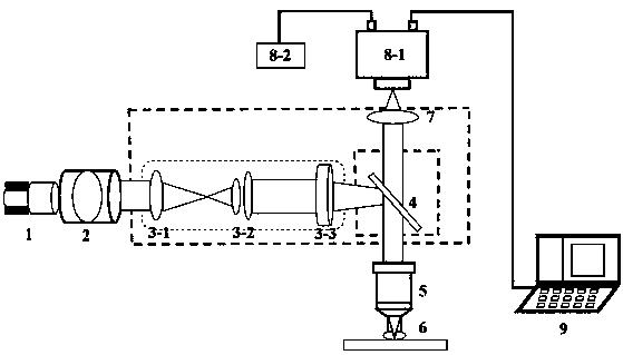
[0019] Such as figure 1 As shown, the present invention is composed of an LED light source, a collimating lens, a microscope epi-illuminator, a dichroic lens, an objective lens, a tube lens, a filter, an InGaAs camera, and a computer.

[0020] The metal radiator 1 is attached to the back of the LED lamp beads for heat dissipation, and then the light source is connected to a tube 2 with a collimating lens inside, and the distance between the LED light source and the collimating lens is adjusted to achieve the best The collimation effect, the beam after collimation has the smallest divergence angle, and the brightness is uniform in the entire spot range, and the diameter of the spot is equivalent to the diameter of the collimating lens (about 50mm). Connect the collimated LED to the rear port of the microscope epi-illuminator through the above-mentioned sleeve, and the collimated light is...


PUM


Abstract

The invention discloses an LED-excited short wavelength infrared fluorescent microscopic imaging system. In the system, laser light emitted by a single-color LED passes through a collimating lens to be collimated firstly and is introduced into a light path of a microscope vertical illumination device, the exciting light is reflected with a dichroic beam combiner and converges onto a back focal plane of a microscopic objective, converging light forms parallel exciting light beams after passing through the objective and illuminates onto a sample, and a fluorescence probe in the sample is excitedto emit fluorescence; and a fluorescence signal passes through the objective, penetrates through the dichroic beam combiner and is finally received by a detection surface of an InGaAs camera to realize photoelectric conversion, and the signal is transmitted to a computer for data processing to obtain an imaged picture or a video. The LED-excited short wavelength infrared fluorescent microscopic imaging system has the remarkable advantages of high signal-to-noise ratio, high magnification, high resolution, capability of real-time imaging, capability of tomography, large penetration depth, small tissue damage and the like.

Description

technical field [0001] The invention belongs to the field of microscopic imaging of applied optics, and is a microscopic imaging system which adopts LED excitation, combines traditional fluorescent microscopic imaging and is applied in the short-wave infrared band. Background technique [0002] Fluorescence microscopy has been widely used in life science research. Fluorescence imaging refers to imaging using fluorescence emitted by biological tissues or structures. Since certain fluorescent dyes can be specifically labeled, fluorescence imaging has the ability to provide specific position information. Microscopic imaging refers to the use of microscopic objective lenses to achieve larger magnification imaging. A typical fluorescence microscopy imaging system includes a light source, a beam splitter, an objective lens, a fluorescent sample, and a photodetector. The excitation light from the light source passes through the illumination optical path and then irradiates the s...

Claims


Application Information

Patent Timeline
IPC IPC(8): G01N21/64G01N21/01
CPCG01N21/01G01N21/64
Inventor 钱骏蔡朝冲
Owner ZHEJIANG UNIV
Why Eureka
Try Eureka