Green fluorescent protein-based nanoparticle, preparation method and application thereof in cell imaging and cell nucleus nucleolar staining

A green fluorescent protein and nanoparticle technology, applied in the field of fluorescent protein-based nanomaterials, can solve complex, expensive, and not commercially available problems, and achieve good biocompatibility, no-cleaning imaging, and cost-effective effects

Active Publication Date: 2018-12-28
JILIN UNIV
View PDF6 Cites 2 Cited by
  • Summary
  • Abstract
  • Description
  • Claims
  • Application Information

AI Technical Summary

Problems solved by technology

However, this process requires a specific antibody, which is often expensive and often not commercially available, and still requires another dye to be bound to the antibody
So, the overall evaluation of this method is complex, expensive
Not conducive to a rapid push for scientific experiments

Method used


Image

  • Green fluorescent protein-based nanoparticle, preparation method and application thereof in cell imaging and cell nucleus nucleolar staining
  • Green fluorescent protein-based nanoparticle, preparation method and application thereof in cell imaging and cell nucleus nucleolar staining
  • Green fluorescent protein-based nanoparticle, preparation method and application thereof in cell imaging and cell nucleus nucleolar staining

Examples


Embodiment 1

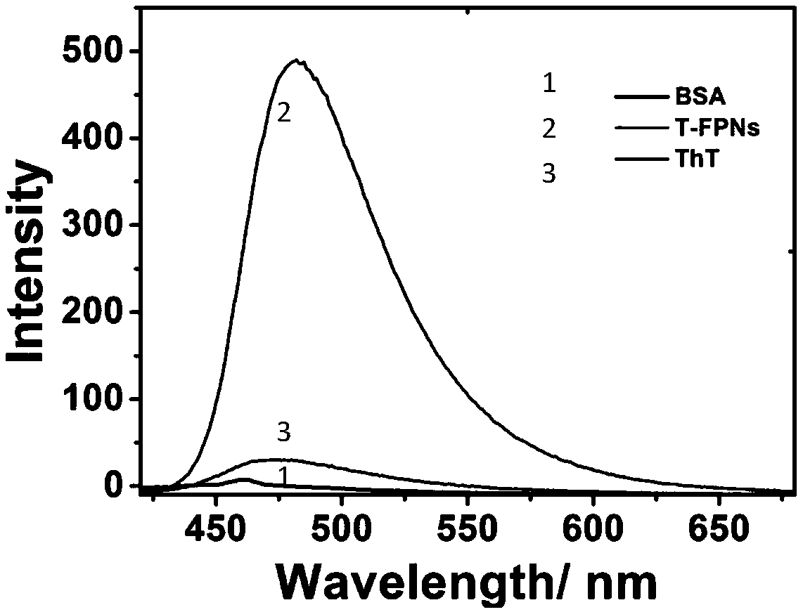
[0016] In 0.8mL deionized water, add 0.1mL of Thioflavin-T aqueous solution with a concentration of 1mol / L as building molecules, add 0.1mL of Bovine Serum Albumin aqueous solution with a concentration of 1mol / L, and the dosage of Thioflavin-T and Bovine Serum Albumin in moles The ratio is 1:1. Then the solution was heated at a temperature of 100° C. for 10 minutes, and finally the solution was cooled to room temperature to obtain a green fluorescent protein-based nanoparticle solution. The concentration of the fluorescent protein-based nanoparticle was 6.4 mg / mL.

Embodiment 2

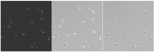
[0018] Saccharomyces cerevisiae cells use YPD (Yeast Extract Peptone Glucose Medium) as the culture medium, and incubate with shaking at 30°C until the OD600 reaches 1.0; take 1 mL of cell culture fluid and centrifuge to collect Saccharomyces cerevisiae cells, and wash the resulting cell strain with fresh YPD three times, then Resuspend the cells in 100 μL of YPD medium containing 0.1 mg / mL green fluorescent protein-based nanoparticles obtained in Example 1, and then put them back into a constant temperature shaker at 30°C and incubate for 6 hours; then take 1 mL of cell culture medium and centrifuge Collect the Saccharomyces cerevisiae cells, and resuspend the resulting cell strains in PBS buffer (pH 7.4) for 3 to 5 times to wash away the medium and fluorescent nanoparticles in vitro as much as possible to avoid background interference during confocal imaging; finally, 2μL The resuspension was dropped on a glass slide, and the cell imaging study was performed using a ZEISS LSM7...

Embodiment 3

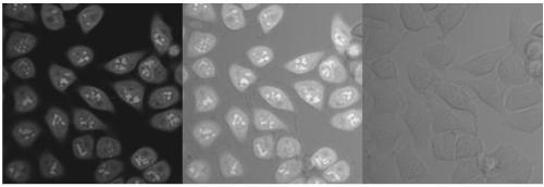
[0020] Inoculate HeLa cells on a cover glass, culture in Dulbecco's modified Eagle medium (DMEM), change the culture medium once every 3 days, and control the incubator environment at 37°C and 5% CO 2 . The steps for staining nucleoli with fluorescent protein-based nanoparticles are as follows. Inoculate the above-mentioned cultured Hela cells in a cell containing 1 mg mL -1 T-FPNs were cultured on DMEM coverslips for 1 hour. After that, use a new cover glass to gently cover the cell-grown cover glass into the imaging chamber, wash twice with Hank's balanced salt solution (HBSS) and keep it in HBSS. Finally, imaging was performed using a ZEISS LSM700 confocal microscope equipped with a 100x oil objective lens (NA1.4) controlled by Zen software, 488nm and 555 lasers. Used to observe the effect of nucleolar staining.


PUM

PropertyMeasurementUnit
concentrationaaaaaaaaaa

Abstract

A green fluorescent protein-based nanoparticle, a preparation method and an application thereof in cell imaging and cell nucleus nucleolar staining belong to the field of fluorescent protein-based nanomaterials. The method includes the steps of adding a concentration thioflavin-T aqueous solution as a building molecule in deionized water, adding a bovine serum albumin or human serum albumin aqueous solution, heating the solution and finally cooling the solution to the room temperature to obtain a green fluorescent protein-based nanoparticle T-FPNs solution. An experiment with Hela cells in yeast cells shows that T-FPNs can be used as bioimaging materials and nucleolar localization dyes. The nanoparticle is suitable for large-scale production because a synthesis method is cost-effective, environmentally friendly, simple and biocompatible. The T-FPNs with excellent light stability can stain nucleoli of live cells and fixed cells nucleoli for fast real-time no-clean imaging.

Description

Technical field [0001] The invention belongs to the field of fluorescent protein-based nanomaterials, and specifically relates to a green fluorescent protein-based nanoparticle, a preparation method and application thereof in cell imaging and nucleolar staining. Background technique [0002] In recent years, various nanomaterials with different functions and properties have made great progress in the design and manufacture. Most of the developed nanomaterials (semiconductor quantum dots, fluorescent carbon dots, luminescent silicon nanoparticles, organic small molecules and organic fluorescent nanoparticles, etc.) are used in bioimaging, drug delivery and other medical diagnosis and treatment applications. But at the same time, the application of these materials has been restricted to varying degrees, such as lack of critical biocompatibility, high toxicity to organisms, complex synthesis routes, high costs, and difficulty in comprehensive promotion. [0003] The nucleolus of the ...

Claims


Application Information

Patent Timeline
Patent Type & Authority Applications(China)
IPC IPC(8): C07K14/47C09K11/06C12Q1/02C12Q1/04
CPCC07K14/47C09K11/06C09K2211/14C12Q1/04G01N33/5005
Inventor 陈志俊李振华
Owner JILIN UNIV
Why Eureka
Try Eureka